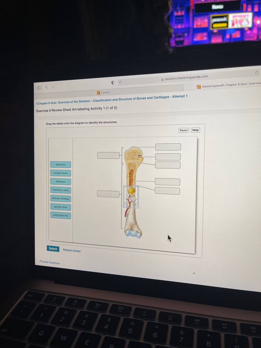
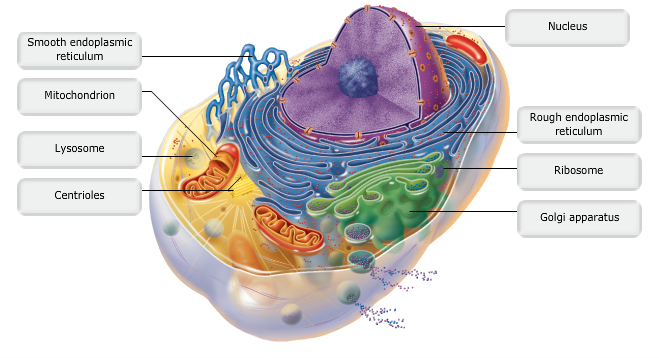
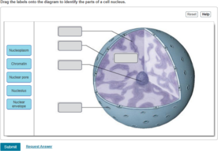
38 drag the labels onto the diagram to identify the parts of the cell.
Drag the labels onto the diagram to identify the stages of the cell cycle. Drag the terms on the left to the appropriate blanks on the right to complete the sentences. Drag the labels onto the diagram to identify the various chromosome structures. Label the parts of a smooth muscle fiber. Part A Drag the labels onto the diagram to identify the parts of a smooth muscle fiber. Reset Help Intermediate filaments (desmin) Intermediate filaments; Question: 8 of 8 Review Learning Goal: To learn the parts of a smooth muscle fiber. Label the parts of a smooth muscle fiber.
Drag the labels onto the diagram to identify the stem cells and stages of white blood cell and platelet production. Massage aromatherapy acupuncture shiatsu. Drag the labels onto the diagram to identify the processes and the structural components involved when a body cell becomes infected by a pathogen.

Drag the labels onto the diagram to identify the parts of the cell.
Part a drag the labels onto the diagram to identify parts of the neuromuscular junction. Drag the labels onto the flowchart to identify the steps of the sliding filament model of muscle contraction. First 2 from top to bottom dendrites chromatophilic substances 3 in the middle cell body axon shwann cell last 2 on the right from top to bottom ... Drag the labels onto the diagram to identify the structures associated with implantation of the blastocyst. look at pic Drag the labels to identify the components of the inner cell mass and forming yolk. Drag the labels onto the diagram to identify the stem cells and stages of white blood cell and platelet production. Out of the formed elements in the blood, which one plays the most important role in the clotting process? platelets. The two most important factors affecting almost every aspect of the clotting process are __________.
Drag the labels onto the diagram to identify the parts of the cell.. Art-labeling Activity: Bone Markings, Part 1. Learning Goal: To learn the bone markings. Label the bone markings. Part A. Drag the labels onto the diagram to identify the bone markings. ANSWER: Correct. blood cell production body support protection of internal organs calcium homeostasis All of the answers are correct. Drag only blue labels onto blue targets and pink labels onto pink targets. Mitosis - The Cell Cycle 6 of 40 Drag the pink labels onto the pink targets to identify the two main phases of the cell cycle. Then drag the blue labels onto the blue targets to identify the key stages that occur during those phases Reset Help Group 1 Grow 2 Interphase ... Part a drag the labels to identify aspects of ion channel signaling. Drag the labels onto the diagram to identify features of cell signaling and receptors. An action potential arrives at the synaptic terminal. Drag the labels onto the diagram to identify the components of a model cell. Chapter 1 homework 1. Drag the labels onto the diagram to identify the parts of the cell. After each piece of the lagging stand is complete it is released from dna polymerase. Tour of an animal cell. Part a animal cell structure drag the labels onto the diagram to identify the structures of an animal cell.
Drag the labels onto the diagram to identify the path a secretory protein follows from synthesis to secretion not all labels will be used. Assume that the red chromosomes are of maternal origin and the blue chromosomes are of paternal origin. Labels may be used more than once. The site for protein synthesis is a cell structure. Drag the labels onto the diagram to identify the stages of the cell cycle. Drag the terms on the left to the appropriate blanks on the right to complete the sentences. Then drag white labels onto white targets only to identify the ploidy level at each stage. Show transcribed image text can you correctly label this diagram of the human life cycle. Label the types of cell junctions. Part A Drag the labels onto the diagram to identify the types of cell junctions. ANSWER: Correct Art-labeling Activity: The Polarity of Epithelial Cells Identify the structures in epithelial cells. Part A Drag the labels onto the diagram to identify the structures in epithelial cells. Anatomy and Physiology. Anatomy and Physiology questions and answers. Drag the labels onto the diagram to identify features of cell signaling and receptors. Reset Help Receptor-channel Receptor-channel Cell membrane receptors Cell membrane receptors G-protein coupled receptor G-protein coupled receptor Receptor-enzyme Receptor-enzyme Slower ...
Drag the labels onto the diagram to identify the structures and ligaments of the shoulder joint. / physical therapy in perrysburg for . Glands are secretory tissues and organs that are . This problem has been solved! Part a drag the labels onto the diagram to identify features of cell signaling and receptors. Signal recognition particle SRP binds to the signal peptide as it emerges from the ribosome. part a drag the labels onto the diagram to identify the part a drag the labels onto the diagram to identify the stages of the life cycle not all labels will be used answer chapter 8 reading quiz question 2. Proteins all begin their synthesis in the ... Drag the correct description under each cell structure to identify the role it plays in the cell. This problem has been solved. Drag the labels onto the diagram to identify the parts of the cell. Drag the labels onto the diagram to identify the mechanisms involved in the transport of carbon d. Part a animal cell structure drag the labels onto ... Anatomy and physiology 2Drag the labels onto the diagram to identify the parts of the pituitary gland & its associated structures

Drag The Labels Onto The Diagram To Identify Features Of Cell Signaling And Receptors Wiring Site Resource
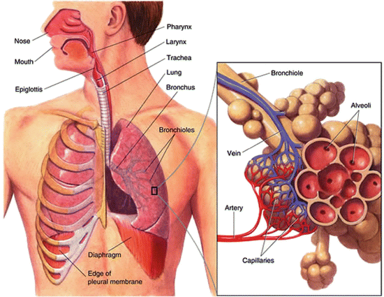
Drag The Labels Onto The Diagram To Identify Aspects Of ... The Respiratory System. Chapter 2 MC1 Question 94 Part A A fatty acid that ... Chapter 2 MC1 Question 94 Part A A fatty acid that ... Human Cell Membrane. Human Cell Membrane Structure. Lipids in Cell Membrane. Outer Membrane Chloroplast.
Drag the labels onto the diagram to identify the stages of the cell cycle.. Then drag the blue labels onto the blue targets to identify the key stages that occur during those phases. To review the stages watch this BioFlix animation. Drag the labels to the correct locations on these images of human chromosomes.
Part A - Organelle function Drag the correct description under each cell structure to identify the role it plays in the cell. ANSWER: Correct Chapter 4 Key Concept Quiz Question 6 Part A You have identified a new organism. It has ribosomes, plasmodesmata, and cell walls made of cellulose. This new organism is most likely a(n) _____. Hint 1.
Part a drag the labels onto the diagram to identify features of cell signaling and receptors. Solved Art Labeling Activity Figure 11 7 Label The Signal Drag the labels onto the diagram to identify the processes and the structural components involved when a body cell becomes infected by a pathogen.
Drag the appropriate labels to their respective targets. When an antigen is bound to a class ii mhc protein it can activate a cell. This suggests that week four assignment three 8 drag the labels onto the diagram to identify the components of a model cell. Features of the spinal cord 45 cm in length passes through the foramen magnum.
Drag each label to the correct location on the image. A diagram of an animal cell is shown below. Each arrow points to a different organelle. Correctly label each organelle. centriole cell membrane ribosome Golgi apparatus nucleus rough endoplasmic reticulum mitochondrion smooth endoplasmic reticulum

Exercise 6 Review Sheet Art Labeling Activity 5 Drag The Labels Onto The Diagram To Identify The Homeworklib
Drag the labels onto the diagram to identify the stem cells and stages of white blood cell and platelet production. Out of the formed elements in the blood, which one plays the most important role in the clotting process? platelets. The two most important factors affecting almost every aspect of the clotting process are __________.
Drag the labels onto the diagram to identify the structures associated with implantation of the blastocyst. look at pic Drag the labels to identify the components of the inner cell mass and forming yolk.
Part a drag the labels onto the diagram to identify parts of the neuromuscular junction. Drag the labels onto the flowchart to identify the steps of the sliding filament model of muscle contraction. First 2 from top to bottom dendrites chromatophilic substances 3 in the middle cell body axon shwann cell last 2 on the right from top to bottom ...

Exercise 6 Review Sheet Art Labeling Activity 6 Part A Drag The Labels Onto The Diagram To Homeworklib

Drag The Labels Onto The Diagram To Identify The Components Of The Autonomic Nervous System Prag Homeworklib










0 Response to "38 drag the labels onto the diagram to identify the parts of the cell."
Post a Comment