38 diagram of uterus and bladder
Bladder Anatomy, Function & Diagram | Body Maps Bladder. The bladder, like the stomach, is an expandable saclike organ that contracts when it is empty. The inner lining of the bladder tucks into the folds and expands out to accommodate liquid ... Pelvic Anatomy - Baystate Ob/Gyn Group Pelvic floor muscles - A group of muscles in the pelvis that support and help to control the vagina, uterus, bladder urethra and rectum Bladder - A muscular organ which stores urine Ureters - A pair of tubes, each leading from one of the kidneys, to the bladder Urethra - A short narrow tube ...
Diagram Uterus Stock Illustrations - 1,534 Diagram Uterus ... Download 1,534 Diagram Uterus Stock Illustrations, Vectors & Clipart for FREE or amazingly low rates! New users enjoy 60% OFF. 183,466,165 stock photos online.

Diagram of uterus and bladder
Uterus Diagram Illustrations, Royalty-Free Vector Graphics ... Browse 1,025 uterus diagram stock illustrations and vector graphics available royalty-free, or search for female reproductive system or uterus icon to find more great stock images and vector art. Human anatomy female reproductive system, female reproductive... Female reproductive system. Vector flat illustration uterus diagram stock illustrations. Anatomy, Abdomen and Pelvis, Uterus - StatPearls - NCBI ... The uterus is located between the urinary bladder anteriorly and the rectum posteriorly. The average dimensions of the uterus in an adult female are 8 cm long, 5 cm across, and 4 mm thick. The uterine cavity has an average volume of 80 mL to 200 mL. The uterus subdivides into three segments, namely: the body, the cervix, and the fundus. ... Position | Uterus Anteverted and "V Shaped Uterus with an Empty Bladder The diagram represents a sagittal view of the uterus reflecting an 'V" shaped structure of the uterus and vagina The uterus varies in position and in this case is anteverted, that converts the L shaped structure described to a "V" shaped structure
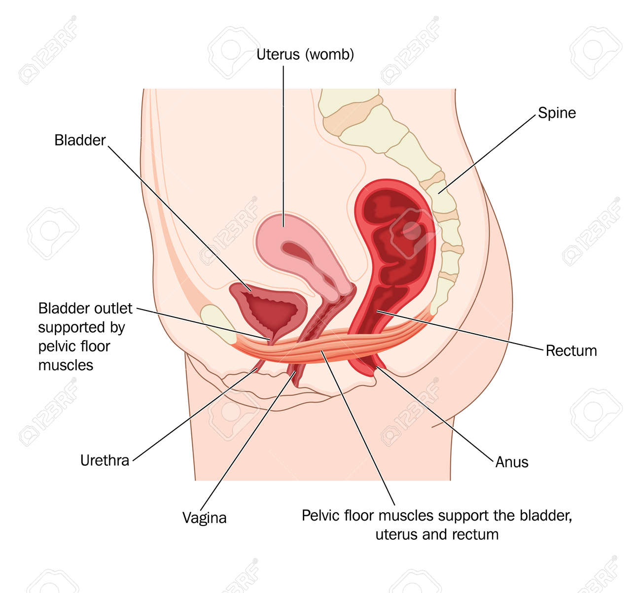
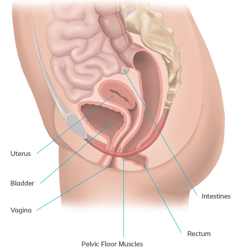
Diagram of uterus and bladder. Female Anatomy - Reproductive System and Vagina Diagram ... Located underneath the pelvis, the pelvic floor not only supports the uterus and vagina, but also the bladder, intestines and rectum - ultimately holding them in place and allowing them to function correctly. It is these muscles that will play a major part in the use and removal of your menstrual cup. Uterus: MedlinePlus Medical Encyclopedia Image The uterus is a hollow muscular organ located in the female pelvis between the bladder and rectum. The ovaries produce the eggs that travel through the fallopian tubes. Once the egg has left the ovary it can be fertilized and implant itself in the lining of the uterus. The main function of the uterus is to nourish the developing fetus prior to ... Transabdominal ultrasound of the uterus. Note the urinary ... Download scientific diagram | Transabdominal ultrasound of the uterus. Note the urinary bladder, uterus, abdominal wall muscles and Pouch of Douglas from publication: Pelvic Ultrasound Simulation ... Uterus Diagram Photos and Premium High Res Pictures ... Browse 229 uterus diagram stock photos and images available, or search for female reproductive system or uterus icon to find more great stock photos and pictures. An anatomical diagram depicts the method of extracting a fetus by reaching into the uterus and adjusting the baby's position.
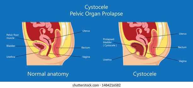
Pelvic Organ Prolapse (dropped bladder, bowel, rectum, uterus) ... Pregnancy, childbirth or extra weight can stretch and weaken muscles that support your pelvic organs. A sheet of muscles and ligaments called the pelvic floor supports the uterus, small bowel, colon and bladder. If pelvic floor muscles are weak, your organs may drop and bulge into the vagina. Bladder (Urinary), Anatomy, Location, Parts and Pictures ... The empty urinary bladder is somewhat tetrahedral in shape - like a three sided pyramid with a triangular base (illustrated in diagram). This gives the bladder one superior surface (top), two inferolateral surfaces (sides) and a posterior surface (back). The external aspect of the superior surface of the bladder is covered by peritoneum. Prolapse of the Uterus, Bladder, Bowel, or Rectum - HERS ... Prolapse of the Uterus, Bladder, Bowel, or Rectum. If you have questions or need a physician referral, please contact HERS at 610-667-7757. Broad bands of uterine ligaments provide structural support to the uterus and pelvis. The uterine ligaments may weaken, stretch, or they can be damaged or severed during surgery. Pics Of Bladder And Uterus Stock Photos, Pictures ... Browse 2,115 pics of bladder and uterus stock photos and images available, or start a new search to explore more stock photos and images. Human internal organ line icon. Minimal vector illustration with simple outline icons as lung, heart, stomach, bone, brain, kidney, skull and other anatomy parts. Editable Stroke.
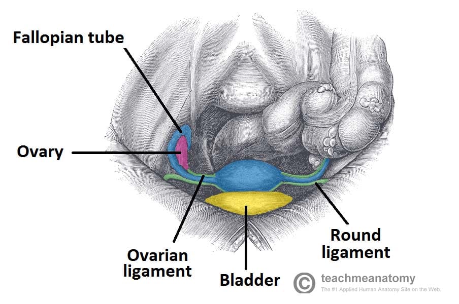
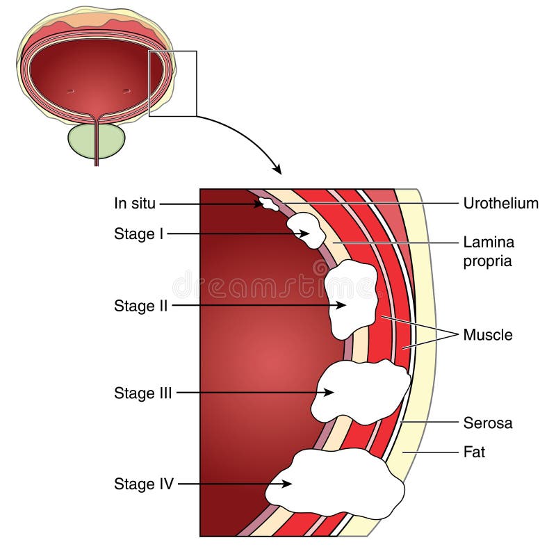
Prolapsed Bladder Pictures, Symptoms, Surgery & Exercises Factors commonly associated with causing a prolapsed bladder are those that weaken the pelvic floor muscles and ligaments that support the bladder, urethra, uterus, and rectum, which can lead to detachment from the ligaments or pelvic bone where the muscles attach: Pregnancy and childbirth: This is the most common cause of a prolapsed bladder. Urinary System, Female, Anatomy: Image Details - NCI ... The uterus is also shown. Anatomy of the female urinary system showing the kidneys, ureters, bladder, and urethra. Urine is made in the renal tubules and collects in the renal pelvis of each kidney. The urine flows from the kidneys through the ureters to the bladder. The urine is stored in the bladder until it leaves the body through the urethra. The female pelvic organs. Bladder, vagina, uterus ... The female pelvic organs: bladder, vagina, uterus, fallopian tube, ovaries. Location and overview Intraoperative view of uterus, bladder, excised VUF tract ... Download scientific diagram | Intraoperative view of uterus, bladder, excised VUF tract, and catheterized ureteral orifices with ureteral stents left for cutaneous externalization. from ...
SMART Imagebase - This medical chart depicts the anatomy of ...
Where Is The Bladder Located In The Human Body Diagram ... The male urethra connects the urinary bladder to the penis. The lower area located inside the vagina named cervix and the widest area named uterus body. Anatomy of the Bladder and Urinary Tract. It is certainly the most widely studied structure the world over. The uterus has two areas. 1-3 It is a hollow organ that is made mostly out of muscle.

Drawing of female pelvis (midsagittal view) shows the anatomy ...
Can Your Uterus or Bladder Fall Out of Your Body? | CIGC April 28, 2020 - Prolapse can be unsettling. Some women may ask "Will my uterus fall out of my vagina if prolapse occurs?" Learn more about pelvic organ prolapse & surgery.

Anatomy location of bladder, vaginal canal, cervix and uterus ...
Biology 12 - The Reproductive System! 17. Label the following diagram and give a function for each labeled part. Name Function UTERUS SITE WHERE EMBRYO DEVELOPS OVIDUCT CONDUCTS EGG TO UTERUS OVARY PRODUCE HORMONES AND RELEASE EGGS VAGINA RECEIVES PENIS AND SERVES AS BIRTH CANAL 18. List 3 functions of estrogen:
Drawing To Show The Pelvic Floor Muscles And Their Support Of ...
Uterine And Bladder Prolapse Guide: Causes, Symptoms and ... The uterus and the bladder are held in their normal positions just above the inside end of the vagina by a "hammock" made up of supportive muscles and ligaments. Wear and tear on these supportive structures in the pelvis can allow the bottom of the uterus, the floor of the bladder or both to sag through the muscle and ligament layers.
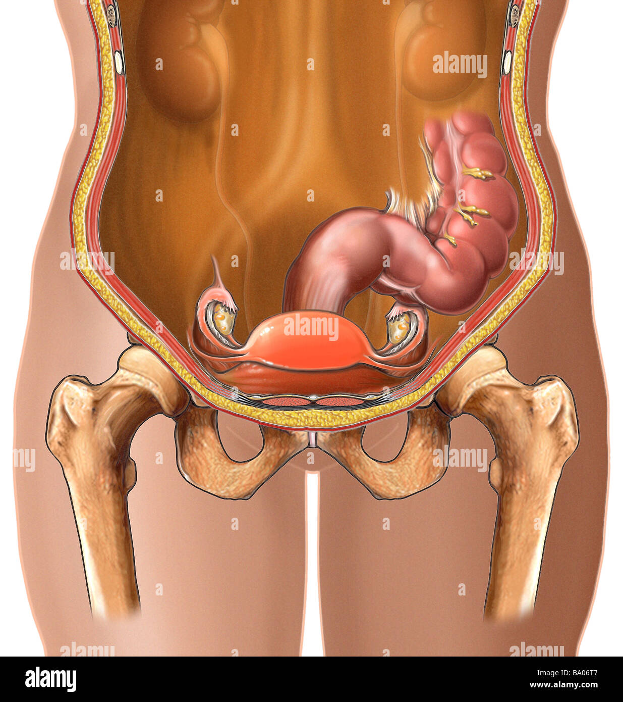
This medical illustration features an anterior view of the ...
Female Pelvis Organs & Inner Muscles Diagram & Function ... January 20, 2018 - Below and in front of the uterus is the bladder. The bladder, also known as the urinary bladder, is an expandable, muscular sac that stores urine. When signaled, the bladder releases urine into the urethra, a tube that carries it out of the body. In women, this tube ends between the clitoris ...

Bladder weakness after birth | Pregnancy Birth and Baby
Anatomy of the Uterus | Female Reproductive Anatomy | Geeky Medics October 22, 2021 - An overview of uterine anatomy, including structure, relations, ligaments, blood supply and innervation.

Drawing To Show The Pelvic Floor Muscles And Their Support Of ...
Anatomy, Abdomen and Pelvis, Female Pelvic Cavity ... The pelvic cavity is a bowl-like structure that sits below the abdominal cavity. The true pelvis, or lesser pelvis, lies below the pelvic brim (Figure 1). This landmark begins at the level of the sacral promontory posteriorly and the pubic symphysis anteriorly. The space below contains the bladder, rectum, and part of the descending colon. In females, the pelvis also houses the uterus ...
The Uterus - Structure - Location - Vasculature - TeachMeAnatomy
Uterine And Bladder Prolapse - Harvard Health July 1, 2019 - What Is It?The uterus and the bladder are held in their normal positions just above the inside end of the vagina by a "hammock" made up of supportive muscles and ligaments. Wear and tear on these supportive structures in the pelvis can allow the bottom of the uterus, the ...

The Female Hip and Pelvis | Musculoskeletal Key
Anatomy location of bladder, vaginal canal, cervix and ... Download scientific diagram | Anatomy location of bladder, vaginal canal, cervix and uterus. CT and the reference US image, the radiation treatment fields ...
Cystocele Treatment - Gynecologic & Reconstructive Surgery
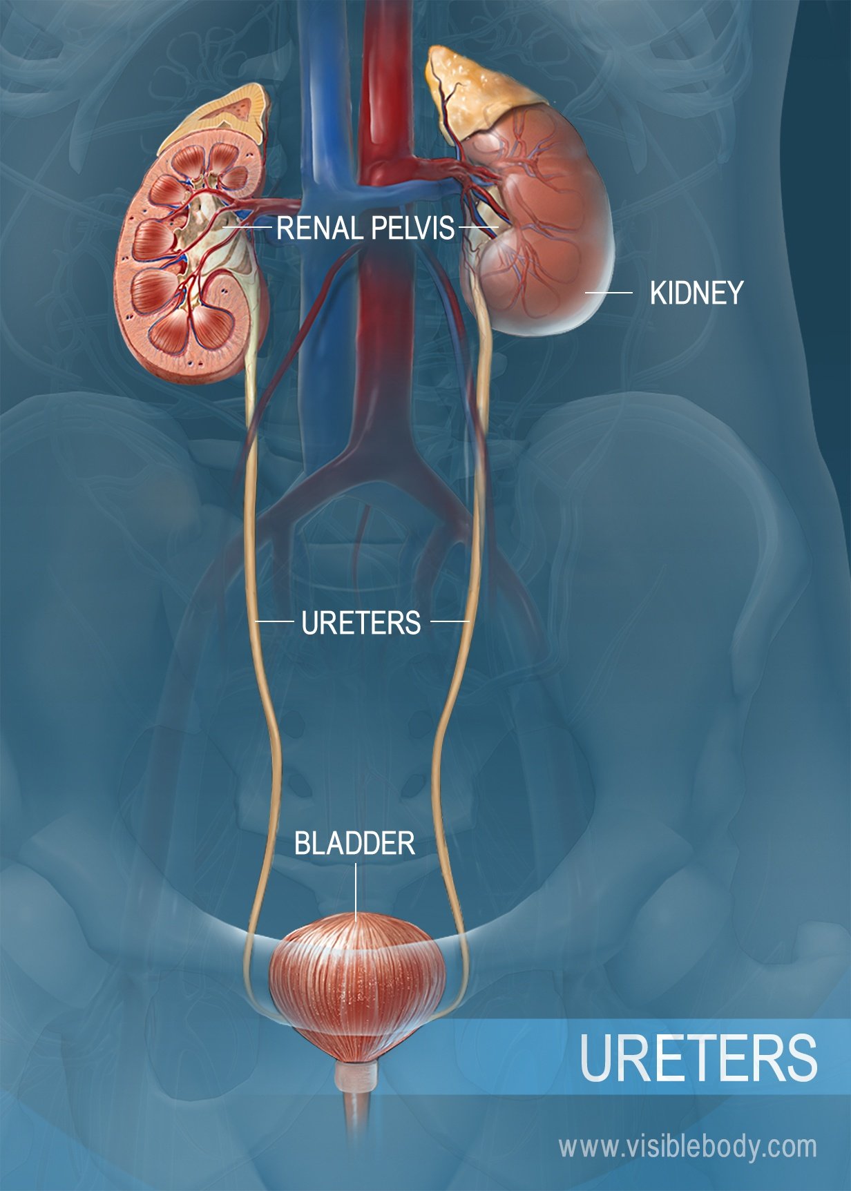
Female reproductive system (urogenital system) anatomy 22 Mar 2006 — It includes the kidneys, ureters, bladder, urethra, and the organs of reproduction – uterus, ovaries, fallopian tubes and vagina.
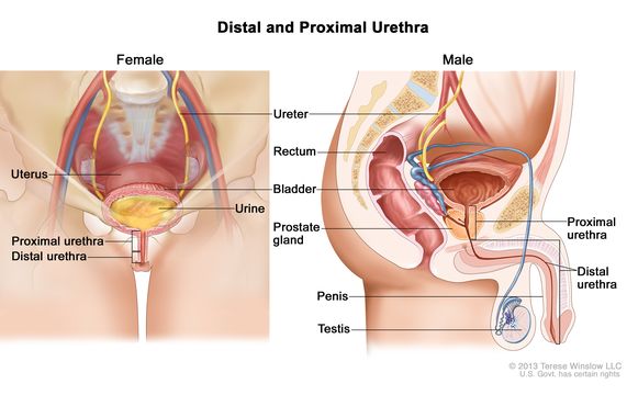
Definition of distal urethra - NCI Dictionary of Cancer Terms ...
Uterus: Anatomy, Function, and Conditions - Verywell Health The uterus, also known as the womb, is the hollow, pear-shaped organ in the female pelvis in which fertilization of an ovary (egg), implantation of the resulting embryo, and development of a baby take place. It is a muscular organ that both stretches exponentially to accommodate a growing fetus and contracts in order to push a baby out during ...

Пин на доске Incontinence Help
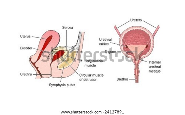
Urinary bladder - Wikipedia The bladder is located below the peritoneal cavity near the pelvic floor and behind the pubic symphysis. In men, it lies in front of the rectum, separated by the recto-vesical pouch, and is supported by fibres of the levator ani and of the prostate gland. In women, it lies in front of the uterus, ...

Fetus I Livmodern Graviditetskvinnor Diagram Gravid Kvinnlig ...
The Uterus - Structure - Location - TeachMeAnatomy This part is structurally and functionally different to the rest of the uterus. See here for more information about the cervix. ... Fig 2 – The three anatomical divisions of the uterus. The exact anatomical location of the uterus varies with the degree of distension of the bladder.
Urinary System Structures
Prolapse & Bladder Weakness | Jean Hailes November 4, 2019 - Uterine and vaginal prolapse. What is it and how is it treated? Learn about types of prolapses, what puts you at a greater risk and how to prevent them.
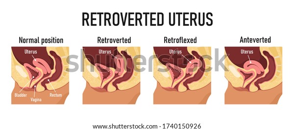
Retroverted Uterus Diagram Variants Uterine Position ...
Uterus bladder Images, Stock Photos & Vectors | Shutterstock Find Uterus bladder stock images in HD and millions of other royalty-free stock ... Hand of woman holding ultrasound image test report from the examination ...
Urinary System, Female, Anatomy: Image Details - NCI Visuals ...
Female reproductive system (urogenital system) information | myVMC March 22, 2006 - It includes the kidneys, ureters, bladder, urethra, and the organs of reproduction – uterus, ovaries, fallopian tubes and vagina. The kidneys are bean shaped organs, which help the body produce urine to get rid of unwanted waste substances. When urine is formed, tubes called ureters transport ...
:max_bytes(150000):strip_icc()/GettyImages-480792143-599ae7596f53ba00115ba667.jpg)
Female Urology and External Sexual Anatomy
Prolapsed Uterus Symptoms, Surgery, Treatment & Exercises August 15, 2018 - When indicated, and in severe cases of prolapse, the uterus can be removed (hysterectomy). During the procedure, the surgeon can also correct the sagging of the vaginal walls, urethra, bladder, or rectum. The surgery may be performed abdominally (through an incision on the abdomen), vaginally ...
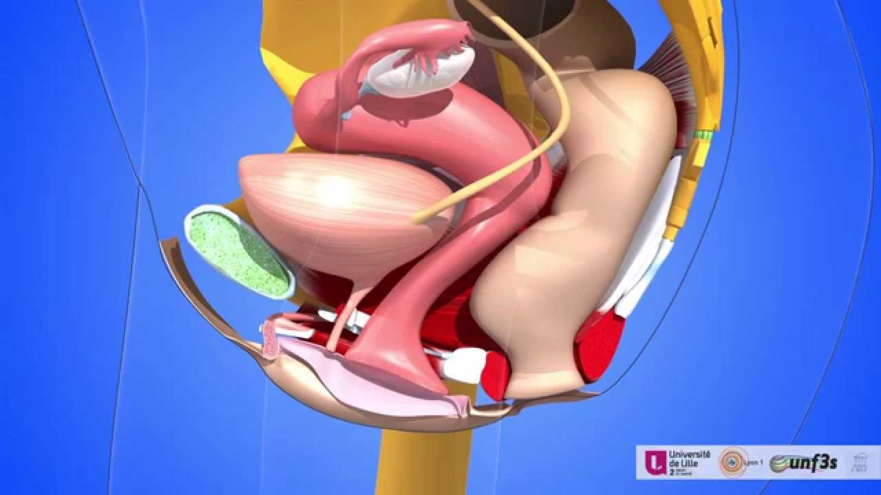
The female pelvic organs. Bladder, vagina, uterus, fallopian tube, ovaries
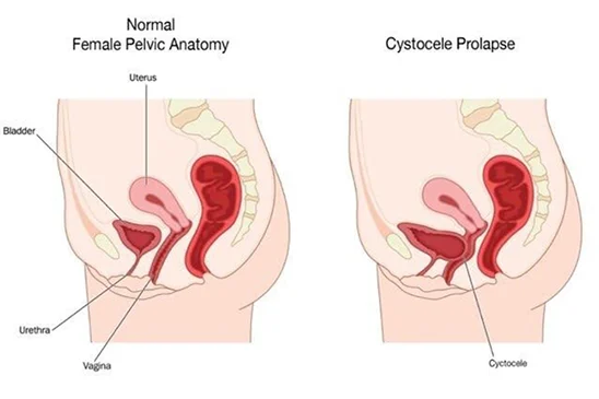
Learn More About Pelvic Organ Prolapse Symptoms ... Cystocele Prolapse: Occurs when the bladder protrudes into the vagina due to the anterior (front) vaginal wall ... Diagram of Uterine Pelvic Organ Prolapse.

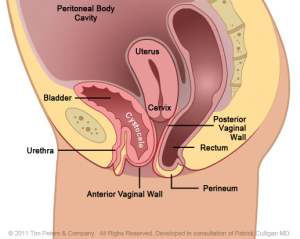
Diagram showing position of bladder, uterus and rectum in a ...
Bladder Diagram Photos and Premium High Res Pictures ... old engraved illustration of the human guts, internal organs - bladder diagram stock pictures, royalty-free photos & images. urine tubes, pelvic viscera, penis, bladder, victorian anatomical drawing - bladder diagram stock illustrations. human reproductive system anatomy - bladder diagram stock illustrations.
Learn More About Pelvic Organ Prolapse Symptoms & Treatments
Uterus and Ovaries - Anatomy Pictures and Information The uterus, also known as the womb, is a hollow, muscular, pear-shaped organ found in the pelvic region of the abdominopelvic cavity. It is located posterior to the urinary bladder and is connected via the cervix to the vagina on its inferior border and the fallopian tubes along its superior end.
Bladder Prolapse Causes, Symptoms and Treatment | Always Discreet
Uterine prolapse - Symptoms and causes - Mayo Clinic Uterine prolapse occurs when pelvic floor muscles and ligaments stretch and weaken and no longer provide enough support for the uterus. As a result, the uterus slips down into or protrudes out of the vagina. Uterine prolapse can occur in women of any age. But it often affects postmenopausal women who've had one or more vaginal deliveries.

Intra operative image showing fetus between the uterus and ...
Urinary bladder & urethra: Anatomy, location, function ... The urinary bladder is found inferior to the peritoneum, sitting on the pelvic floor. In females its inferior surface lays on the pubic symphysis and the posterior wall is in contact with the vagina and uterus.. In males, the inferior surface of the bladder lays over the pubic symphysis and prostate, posteriorly is the distal third of the rectum.Between the posterior surface of the bladder and ...

For patients | pfm medical ag
Diagram of uterus - Healthiack Diagram of uterus 918. Diagram of uterus 925. Diagram of uterus 927. Diagram of uterus 929. Diagram of uterus 930. Diagram of uterus 958. Diagram of uterus 994. Diagram of uterus 1045. Diagram of uterus 1085.
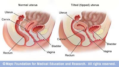
Tipped (tilted) uterus - Mayo Clinic
Female Body Diagram: Parts of a Vagina, Location, Function Vagina: The vagina is a muscular canal that connects the cervix and the uterus, leading to the outside of the body. Parts of the vagina are rich in collagen and elastin, which give it the ability to expand during sexual stimulation and childbirth. Cervix: The cervix is the lower part of the uterus that separates the lower uterus and the vagina and may play a role in lubrication.

Patient education: Pelvic floor muscle exercises (Beyond the ...
Anatomy of the Female Abdomen and Pelvis, Cut-away View ... This medical exhibit diagram illustrates the anatomy of the female abdomen and pelvis from an anterior (front) cut-away view, showing elements of the digestive system. The liver, stomach, and abdominal contents are clearly identified and labeled, including the cecum, ascending colon, transverse colon, descending colon, and small intestine. The image also shows the pelvis, uterus, and urinary ...
Definition of reproductive system - NCI Dictionary of Cancer ...
Parts | Uterus The diagram represents a sagittal view of the uterus reflecting an 'L" shaped structure of the uterus vagina, and the internal cavity. The parts that the tract traverses includes the vagina at the downstream end which now is a significantly (pink), cervix (royal blue), lower uterine segment (light blue), the body (lighter blue), and the ...
Bladder Anatomy and Relation To Uterus Stock Vector ...
Position | Uterus Anteverted and "V Shaped Uterus with an Empty Bladder The diagram represents a sagittal view of the uterus reflecting an 'V" shaped structure of the uterus and vagina The uterus varies in position and in this case is anteverted, that converts the L shaped structure described to a "V" shaped structure

Female pelvic area with labels for the cervix, vagina ...
Anatomy, Abdomen and Pelvis, Uterus - StatPearls - NCBI ... The uterus is located between the urinary bladder anteriorly and the rectum posteriorly. The average dimensions of the uterus in an adult female are 8 cm long, 5 cm across, and 4 mm thick. The uterine cavity has an average volume of 80 mL to 200 mL. The uterus subdivides into three segments, namely: the body, the cervix, and the fundus. ...

Anatomy of the Uterus | Ovaries | 3D Anatomy Tutorial
Uterus Diagram Illustrations, Royalty-Free Vector Graphics ... Browse 1,025 uterus diagram stock illustrations and vector graphics available royalty-free, or search for female reproductive system or uterus icon to find more great stock images and vector art. Human anatomy female reproductive system, female reproductive... Female reproductive system. Vector flat illustration uterus diagram stock illustrations.
Bladder Anatomy Relation Uterus Labeled Stockvektor ...
Rectocele Bilder, stockfoton och vektorer med | Shutterstock
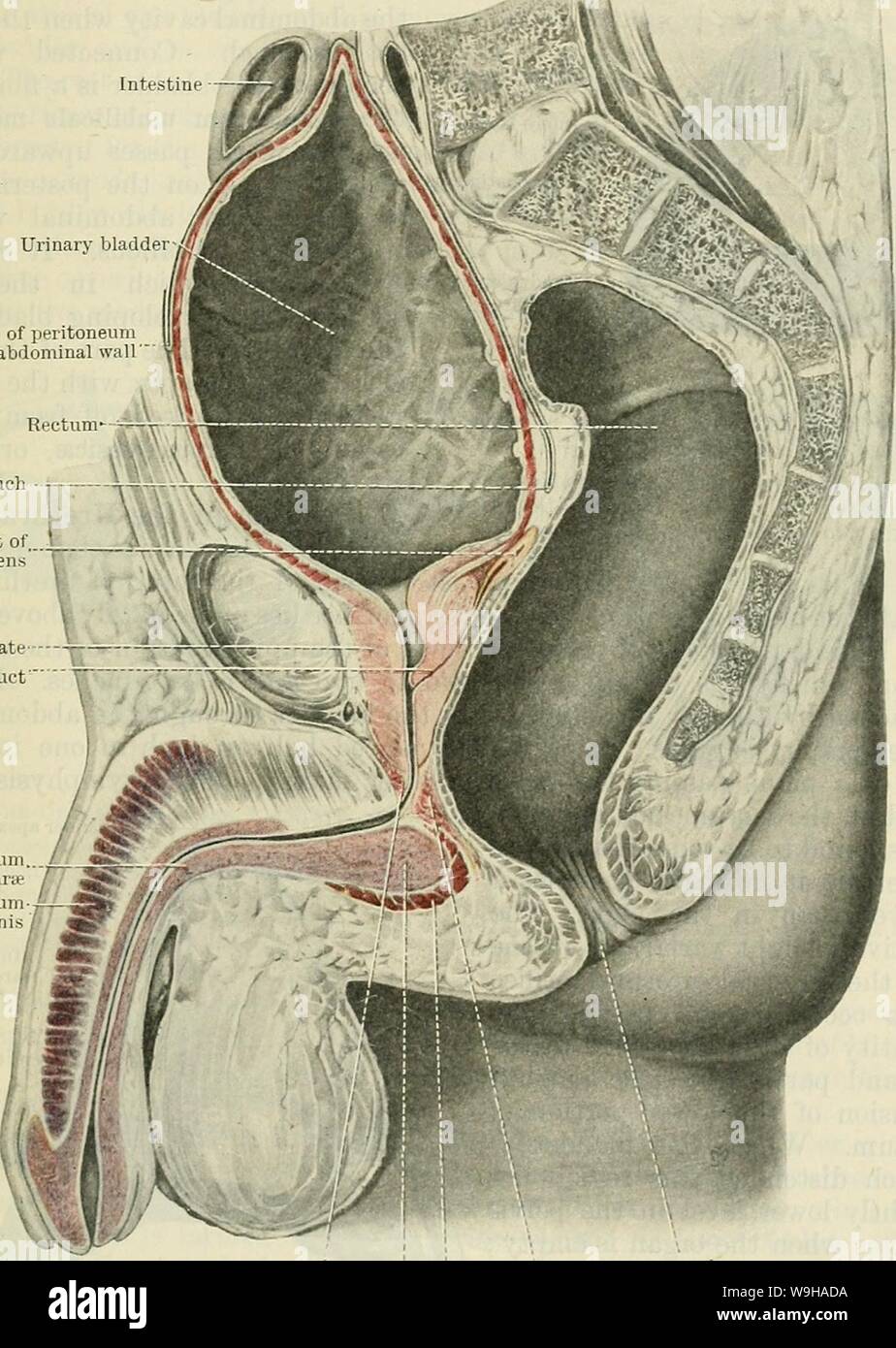
Archive image from page 1306 of Cunningham's Text-book of ...

Female Anatomy Diagram Stock Illustration - Download Image ...

Anatomy of the female pelvis with relations of the urinary ...

Basic Anatomy: The Urinary System | Embarrassing Problems

pelvic spaces, gyn Diagram | Quizlet

Urinary bladder - Wikipedia
An illustrated cross-sectional view of the uterus and bladder ...
0 Response to "38 diagram of uterus and bladder"
Post a Comment