38 ventral body cavity diagram
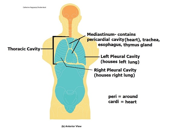
Pelvic cavity - Wikipedia The pelvic cavity is a body cavity that is bounded by the bones of the pelvis. Its oblique roof is the pelvic inlet (the superior opening of the pelvis). Its lower boundary is the pelvic floor. The pelvic cavity primarily contains the reproductive organs, urinary bladder, distal ureters, proximal urethra, terminal sigmoid colon, rectum, and ... Mapping the Body | Boundless Anatomy and Physiology The thoracic cavity is lined by two types of mesothelium, a type of membrane tissue that lines the ventral cavity: the pleura lining of the lungs, and the pericadium lining of the heart. Abdominopelvic. The abdominoplevic cavity is the posterior ventral body cavity found beneath the thoracic cavity and diaphragm.
Body Cavities Diagram | Quizlet ventral body cavity (Anterior View) contains all the structures within the chest and abdomen. Ventral Body Cavities (Lateral View) contains all the structures within the chest and abdomen. Recommended textbook explanations. Introduction to Anatomy and Physiology Michelle Provost-Craig, Susan J. Hall, William C. Rose.
Ventral body cavity diagram
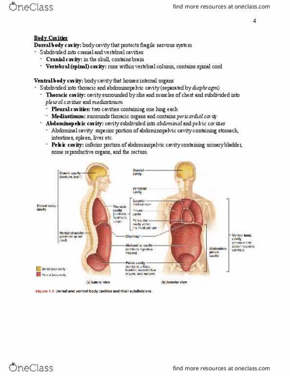
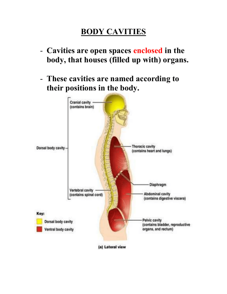
What organs are in ventral cavity? - TreeHozz.com Jun 30, 2020 · The ventral body cavity is a human body cavity that is in the anterior (front) aspect of the human body. It is made up of the thoracic cavity , and the abdominopelvic cavity . The abdominal cavity contains digestive organs, the pelvic cavity contains the urinary bladder, internal reproductive organs, and rectum. Imaging the oral cavity: key concepts for the radiologist The oral cavity is a challenging area for radiological diagnosis. Soft-tissue, glandular structures and osseous relations are in close proximity and a sound understanding of radiological anatomy and common pathways of disease spread is required. In this ... PDF Chap1-anatomical terminology [Compatibility Body organization 1. Body cavities – hollow spaces within the human body that contain internal organs. a) The dorsal cavity: located toward the back of the body, is divided into the cranial cavity (which holds the brain) and vertebral or spinal cavity (which holds the spinal cord). b) The ventral cavity: located toward the front of the body, is
Ventral body cavity diagram. PDF Anatomy & Physiology Cat Muscles Ventral view 1 Ventral view of the superficial and deep musculature of the cat. Rectus abdomnis Internal oblique Stemomastoideus Pectora/is major Pectora/is minor External oblique Linea alba Sanofius Gracifus . O o o o O õ o m o z O c z o o o m N c o o C > m õ C m c m O o > o CD x m > o o CD o . SUPRASPINATUS BRACHIALIS Cow Anatomy - External Body Parts and Internal Organs with ... Jul 28, 2021 · Here in this article, first, I will show you the different body parts of a cow with a labeled diagram. Then I will discuss the anatomy of some vital internal organs of a cow . You should know the details of bones, muscles, digestive organs like stomach, respiratory organs, nerves, and veins from a cow. BLANK ventral body cavity diagram - | Course Hero BLANK ventral body cavity diagram -. School California State University, Long Beach. Course Title BIO 208. Type. Homework Help. Uploaded By cindyaboud07. Pages 1. This preview shows page 1 out of 1 page. View full document. Body Cavities - Simple Anatomy and Physiology Ventral Cavity The ventral cavity is located at the front of the trunk and contains the thoracic cavity and the abdominopelvic cavity. The thoracic cavity and abdominopelvic cavity are separated by the diaphragm, which is a thin skeletal muscle. Thoracic cavity : located in the chest (upper part of the trunk) and contains the heart and the lungs.
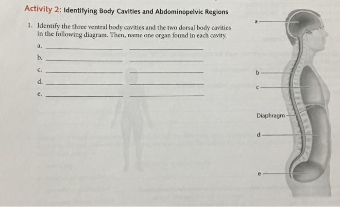
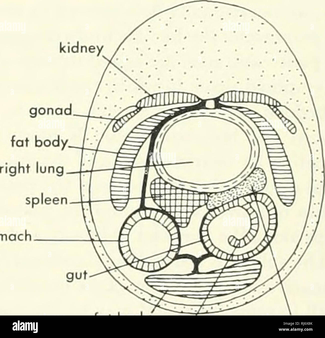
Ventral Body Cavity | Subdivisions, Organs, & Diagram ... The ventral cavity is an example of one type of body cavity, a fluid-filled space that encloses and protects the body's internal organs. The ventral cavity is further subdivided into the thoracic ... Body Cavities and Organs with Labeled Diagram - The Major ... Body cavities diagram from an animal. You already got a different diagram of the body cavity from an animal. Here, I will show you again some of the cavities in one diagram. But, if you need more updated diagrams on the body cavity, you may join anatomy learner on social media. Solved Dorsal Ventral Activity 2: Identifying Body ... Identify the three ventral body cavities and the two dorsal body cavities shown in the following diagram a b. d. e. Diaphragm 2. In which specific body cavity is each of the following organs located? a urinary bladder e spleen b. lungs 1. spinal cord: c. brain: Besophagus d. stomach h. liver 3. The spleen is located in which abdominopelvic ... Beautiful Body Cavities Diagram Unlabeled - Glaucoma Template Search and use 100s of blank body diagram human unlabeled clip arts and images all free. The ventral body chamber that contains the abdominal cavity primarily digestive system and the pelvic cavity primarily reproductive system.
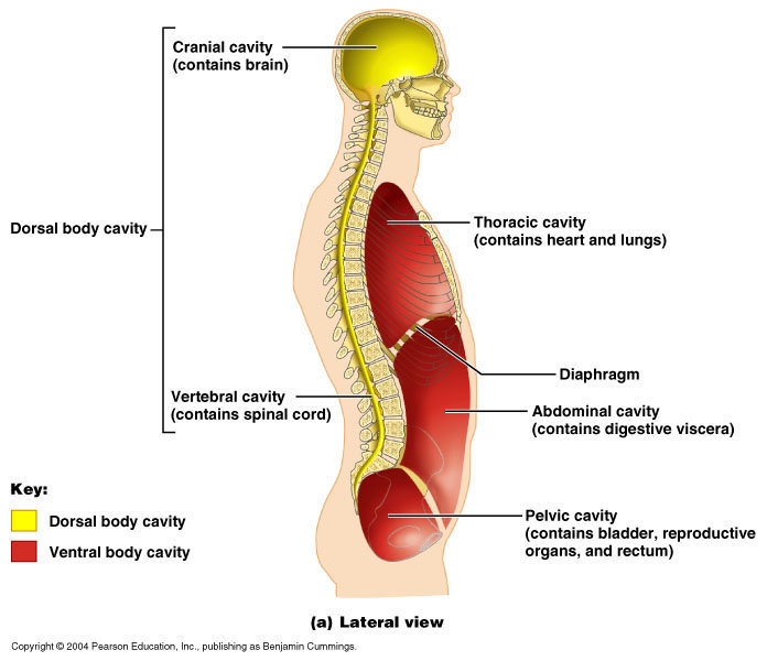
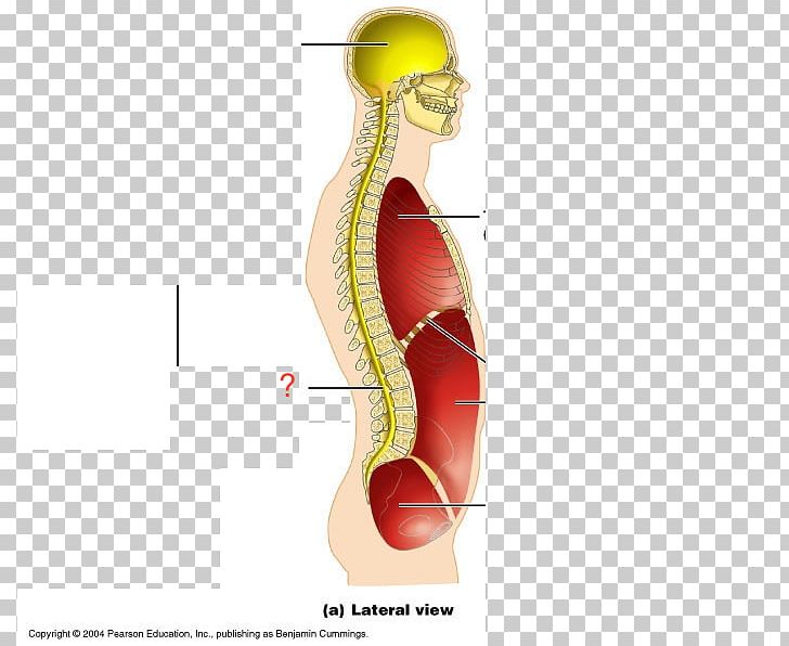
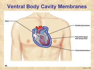
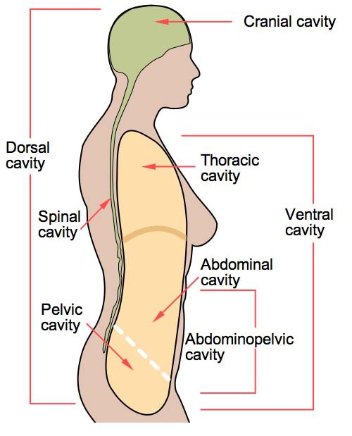
PDF Livingston Public Schools / LPS Homepage Ventral body cavity (a) Lateral view (contains heart and lungs) —Diaphragm Abdominal cavity contains digestive viscera) cavitv (contains bladder, reproductive Rorgans and rectum) Figure 1.9a . Body Cavities Dorsal cavity protects the nervous system, and is divided into two subdivisions Human Nervous System Structure and Functions ... - Bodytomy Diagrams! They remind me of school textbooks which used to have plenty of them, providing a visual aid to understanding difficult subjects. This article explains the nervous system function and structure with the help of a human nervous system diagram and gives you that erstwhile 'textbook feel'. Solved 4. A bullet that lodges in the heart would: be ... PRE-LAB Activity 2: Using Anatomical Terminology to Describe Organ Locations 1. The dorsal body cavity is subdivided into the cavity and the 2. The ventral body cavity is subdivided into the cavity and the 3. Mark each of the following statements as either true (T) or false (F). a. The heart is lateral to the lungs. b. The wrist is distal to ... Body cavities and membranes : Anatomy & Physiology Membranes in the Ventral body cavity. The walls of the ventral body cavity and outer covering of its organs contain a thin covering called the serosa (also called serous membrane). It is a double-layered membrane made up of two parts called the "parietal serosa" (lines the cavity walls) and "visceral serosa" (covers organs in the cavity).
Which body cavities contain the central nervous system ...
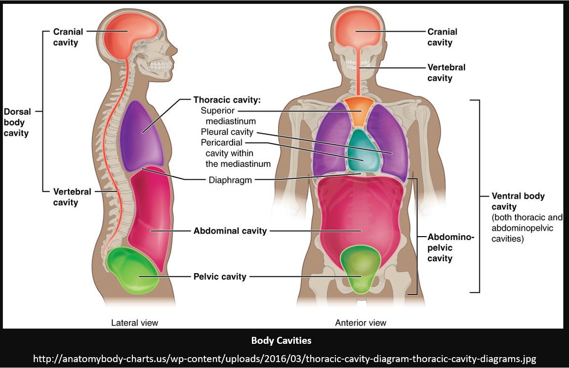
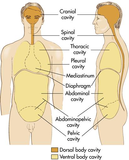
The Human Body Cavities - Lumen Learning Cranial cavity-the space occupied by the brain, enclosed by the skull bones. Spinal cavity-the space occupied by the spinal cord enclosed by the vertebrae column making up the backbone. The spinal cavity is continuous with the cranial cavity. Ventral body cavity-the thoracic cavity, the abdominal cavity, and the pelvic cavity in combination.

Clinical Anatomy & Operative Surgery - Body Cavities | Facebook
Ventral Cavity Diagram - biology 156, lab 1 digestive ... Ventral Cavity Diagram - 16 images - ppt body planes directions and cavities powerpoint, ppt cat dissection powerpoint presentation free, clam dissection biology junction, fetal pig anatomy oral cavity image license carlson,
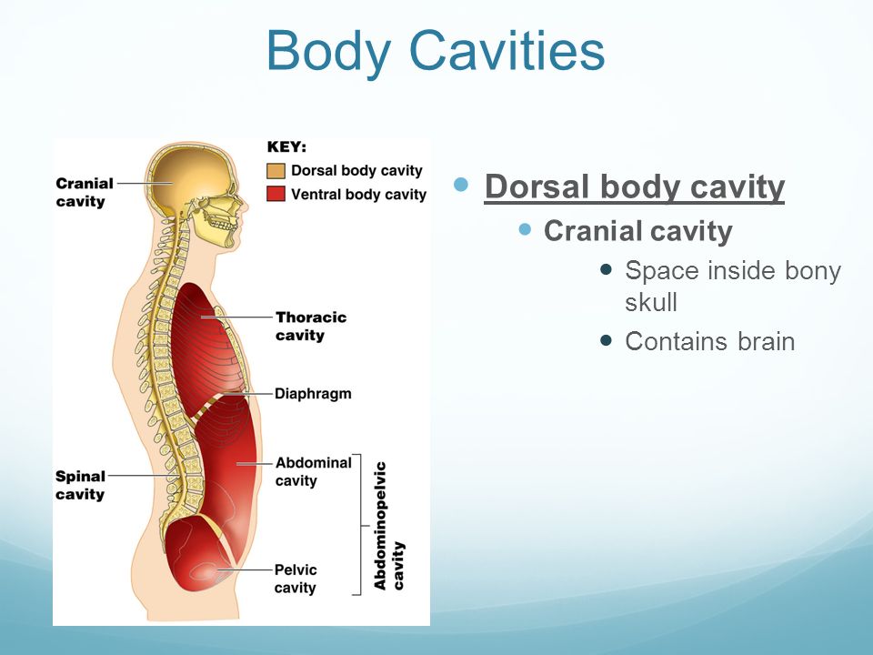
Body Cavities. Dorsal body cavity Cranial cavity Space inside ...
Dissection of Rat (With Diagram) | Zoology The anterior, narrow, looped region opening in the body cavity through a ciliated funnel, facing the ovary. b. Uterus: The thick walled, dilated portion following the fallopian tube: Vagina: The two uteri join to form a median vagina. It is a muscular passage, ventral to the rectum but dorsal to the urethra.

Body Cavities, Illustration - Stock Image - C050/4480 ...
Body Cavities and Membranes: Labeled Diagram, Definitions You can see how the ventral cavity is situated in front of or anterior to the dorsal cavity. The ventral cavity houses the contents within the chest, abdomen, and pelvis. Body Cavities Labeled Diagram: The ventral cavity is located in the front of the body (red/star) and houses the organs/structures of the chest, abdomen, and pelvis. Cranial Cavity
File:Body Cavities labeled.png - Wikimedia Commons
Anatomical terms of location - Wikipedia Dorsal and ventral. These two terms, used in anatomy and embryology, describe something at the back (dorsal) or front/belly (ventral) of an organism. The dorsal (from Latin dorsum 'back') surface of an organism refers to the back, or upper side, of an organism. If talking about the skull, the dorsal side is the top.

Body Cavities - Course Hero
Fetal Pig Dissection Lab - Humble Independent School District Follow the large intestine (colon) to the rectum. This lies in the dorsal wall of the abdominal cavity and is the straight end portion of the large intestine. Water is absorbed by the body in the large intestine. Waste material stored in the rectum leaves the body through the anus.
Solved Identify the three ventral body cavities and the two ...
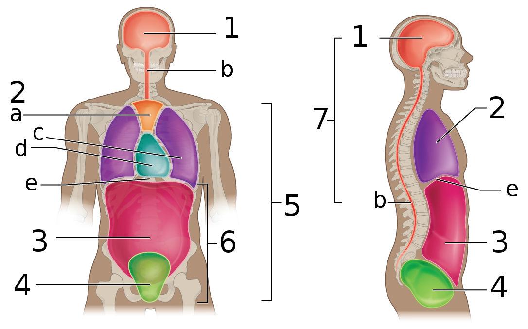
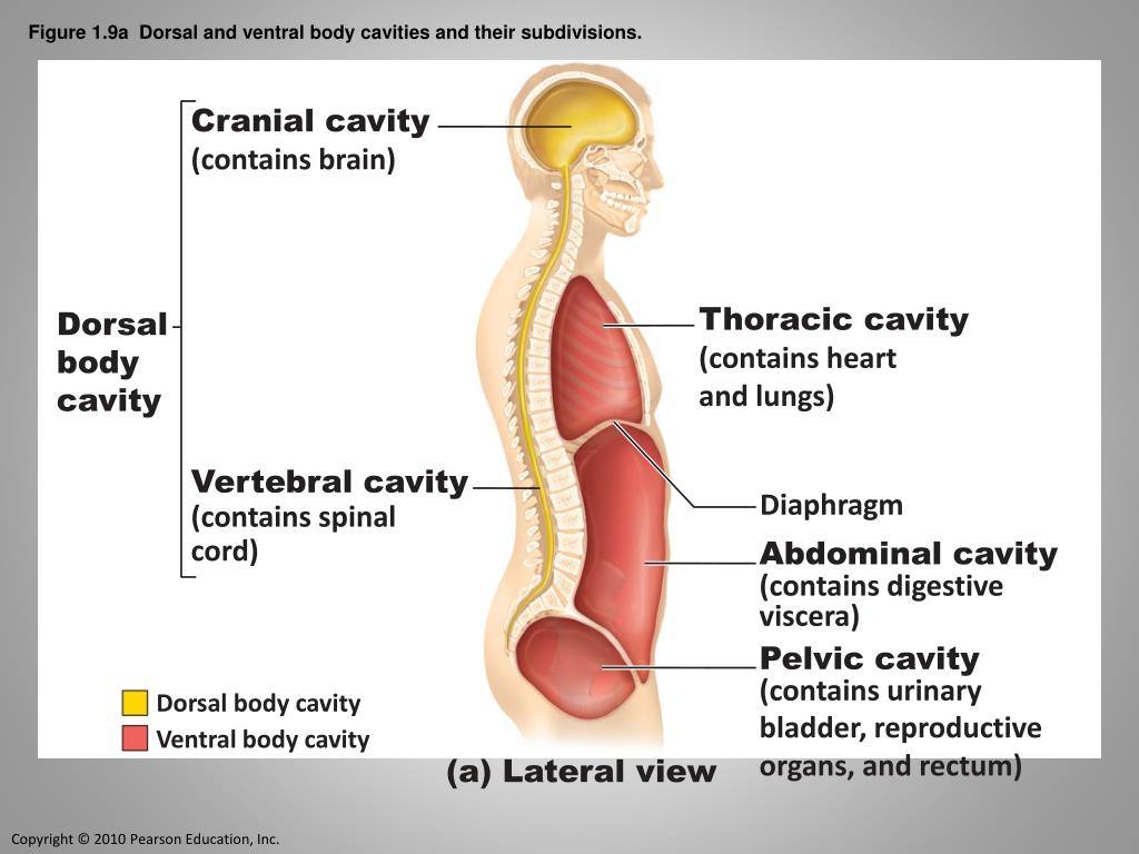
Identify the three ventral body cavities and the two ... Identify the three ventral body cavities and the two. 1. Identify the three ventral body cavities and the two dorsal body cavities in the following diagram. Then, name one organ found in each cavity. a. cranial brain b. vertebral spinal cord c. thoracic heart d. abdominal stomach e. pelvic bladder. 2.
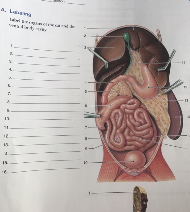
Solved Label the organs of the cat and the ventral body ...
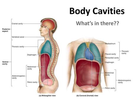
Body Cavities and Membranes - Registered Nurse RN The ventral body cavity is the larger cavity located toward the front of the body, and it contains our visceral organs (or guts!). Remember: ventral contains the viscera! The ventral cavity can also be divided into two main parts: the thoracic cavity and abdominopelvic cavity, which are separated by the diaphragm.
Body Cavities and Membranes – Anatomy and Physiology Notes
31 Body Cavities Label - Labels For Your Ideas Start studying body cavities labeling. The dorsal body cavity protects organs of the nervous system and has two subdivisions. Anterior portion of the torso. Bones of the cranial portion of the skull and vertebral column toward the posterior dorsal side of the body cranial cavity. The spinal cavity is continuous with the cranial cavity.

DOC) Cavidades corporales | Andrea Murrieta Alvarez ...
PDF Diagrams Ch. 1 Body Organization Anatomy & Physiology Ch. 1 Body Organization Diagrams . Body Planes A B C. Regional Terms . Body Cavities . ... .Ventral body cavity (a) Lateral view Thoracic cavity (contains heart and lungs) Cranial cavity Vertebral cavity Superior mediastinum Lung and pleural cavity Mediastinum, with heart and pericardial

Dorsal and Ventral Body Cavities, Lateral View Diagram | Quizlet
PDF Body regions, Major body Cavities - Sinoe Medical Association Dorsal Body Cavity which houses Cavities the CNS: brain and spinal cord 1). Cranial Cavity 2). Vertebral or spinal cavity • • B). Ventral Body Cavity • which houses all other internal body organs 1). Thoracic: • a). Pleural Cavities • b). Pericardial Cavity • c). Mediastinum 2). Abdominopelvic Cavity • a). Abdominal Cavity • b ...
Ch. 1 “An Introduction to the Human Body
Dorsal and Ventral: What Are They, Differences ... - Osmosis On the anterior side of the body, the ventral cavity is made up of the thoracic cavity, abdominal cavity, and pelvic cavity. The thoracic cavity contains the heart, lungs, breast tissue, thymus gland, and blood vessels. Inside the abdominal cavity are the stomach, liver, gallbladder, pancreas, small intestine, colon, appendix, and kidneys.

Chapter 1H Body Cavities
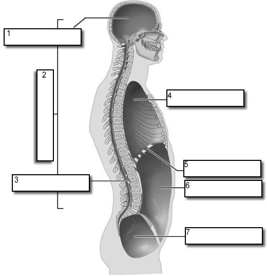
Body Cavities Labeling - The Biology Corner Front View: 1. Cranial Cavity 2. Vertebral Canal 3. Mediastinum 4. Pleural Cavity 5. Pericardial Cavity 6. Diaphragm 7. Abdominal Cavity 8. Pelvic Cavity 9. Abdominopelvic Cavity 10. Ventral Cavity . Side View: 1. Cranial Cavity 2. Dorsal Cavity 3. Vertebral Canal 4. Thoracic Cavity 5. Diaphragm 6. Abdominal Cavity 7. Pelvic Cavity

What Are the Different Types of Human Body Cavities?
Physiology: Human (Ventral) Body Cavities Quiz a body cavity is any fluid filled space in a multicellular organism. However, the term usually refers to the space, located between an animal's outer covering (epidermis) and the outer lining of the gut cavity, where internal organs develop. This quiz has tags. Click on the tags below to find other quizzes on the same subject.

Ventral Body Cavity - Physiology - AmeriCorps Health
Body Cavities Diagram | Quizlet Start studying Body Cavities. Learn vocabulary, terms, and more with flashcards, games, and other study tools.

Ventral body cavity - wikidoc
Reproductive System Review Guide Diagram Answer Key Discover the body's cavities and organs found in the ventral cavity, where it is located, and see a ventral body cavity diagram. Ventral Body Cavity | Subdivisions, Organs, & Diagram 2 days ago · The female sex organs consist of both internal and external genitalia.
Chordate morphology. Morphology (Animals); Chordata ...
Ventral Cavity - Definition and Function | Biology Dictionary The human ventral cavity is divided into two main parts, the thoracic cavity and the abdominopelvic cavity. The thoracic cavity is further divided into separate parts. Two pleural cavities, the left and right, hold the lungs. A central membrane, the mediastinum, divides these two chambers. The heart sits within the pericardial cavity.

1.4E: Body Cavities - Medicine LibreTexts
Medical Terminology Whole Body Medical Terms - GlobalRPH Apr 05, 2021 · Ventral . Ventr/o = belly; equivalent to the anterior or front side of the body-al = pertaining to. The ventral side of the body includes the chest, abdomen, shins, palms, and soles. The ventral cavity in the human body is a fluid-filled space at the anterior side, housing visceral organs. Dorsal

Body cavities and membranes | Human anatomy and physiology ...
Ventral Body Cavity: Definition, Subdivisions & Organs ... The ventral cavity is the front, or belly, cavity of the body. It's divided into the thoracic cavity, which houses the esophagus, trachea, heart, and lungs, and the abdominopelvic cavity, of which ...
BLG 10A/B Study Guide - Fall 2017, Final - Pelvic Cavity ...
PDF Chap1-anatomical terminology [Compatibility Body organization 1. Body cavities – hollow spaces within the human body that contain internal organs. a) The dorsal cavity: located toward the back of the body, is divided into the cranial cavity (which holds the brain) and vertebral or spinal cavity (which holds the spinal cord). b) The ventral cavity: located toward the front of the body, is
Human Body Human Anatomy Ventral Body Cavity PNG, Clipart ...
Imaging the oral cavity: key concepts for the radiologist The oral cavity is a challenging area for radiological diagnosis. Soft-tissue, glandular structures and osseous relations are in close proximity and a sound understanding of radiological anatomy and common pathways of disease spread is required. In this ...
Body Cavities Labeling
What organs are in ventral cavity? - TreeHozz.com Jun 30, 2020 · The ventral body cavity is a human body cavity that is in the anterior (front) aspect of the human body. It is made up of the thoracic cavity , and the abdominopelvic cavity . The abdominal cavity contains digestive organs, the pelvic cavity contains the urinary bladder, internal reproductive organs, and rectum.
Body Cavities and Membranes - SCIENTIST CINDY

Body Cavities/Systems | Medicine Quiz - Quizizz

Ventral Body Cavity Diagram | Quizlet
Body Cavities and Regions. Body Cavities Body Cavities: areas ...
1. Introduction to anatomy and physiology | Nurse Key

Body anatomy, Thoracic cavity, Body

Preparing for A&P: Basic Science and Biology, 1st Edition ...
body cavities - Winston Knoll Collegiate

PPT - The Human Body: An Orientation PowerPoint Presentation ...
Lab 1 the human body
PPT - Language of Anatomy PowerPoint Presentation, free ...

Body cavity - Wikipedia
Anatomical Terms & Meaning: Anatomy Regions, Planes, Areas ...

Dorsal and Ventral Body Cavities Diagram | Quizlet

Dorsal and Ventral Body Cavities · Open Educational Resource ...

File:Body Cavities Frontal view.jpg - Wikimedia Commons
0 Response to "38 ventral body cavity diagram"
Post a Comment