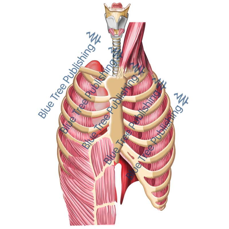
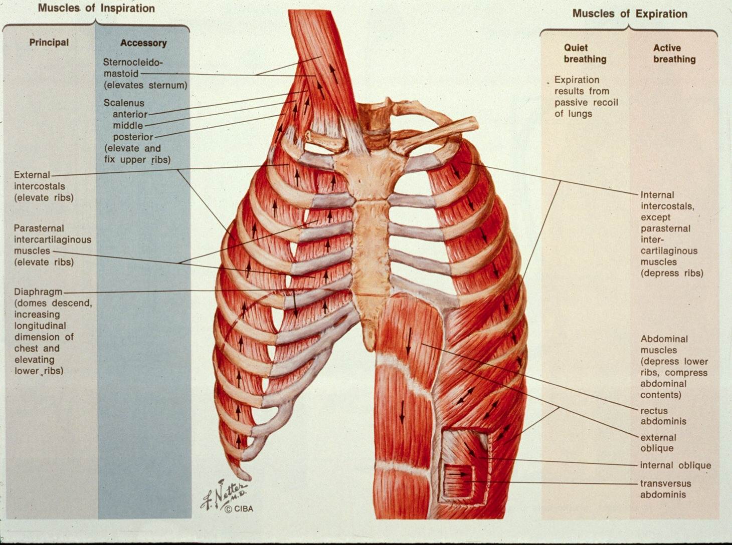
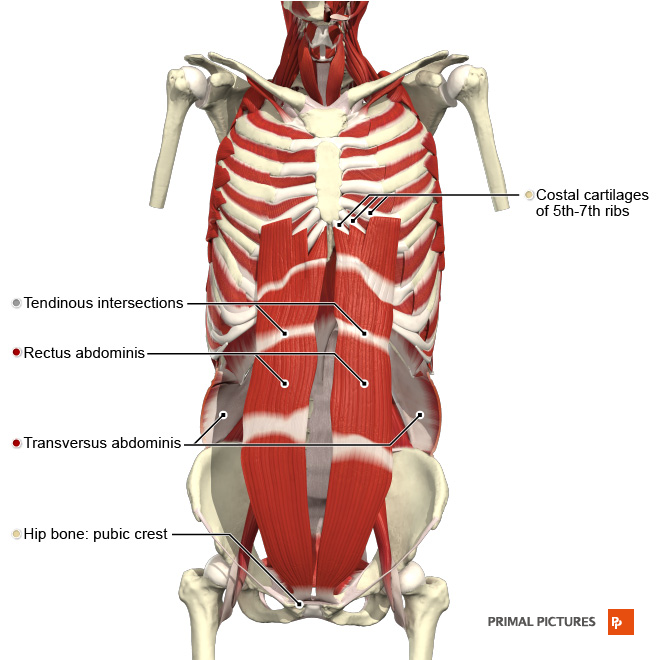
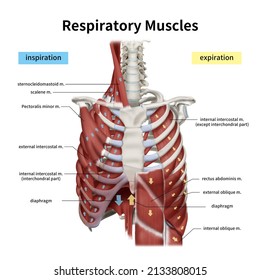
39 muscles of respiration diagram
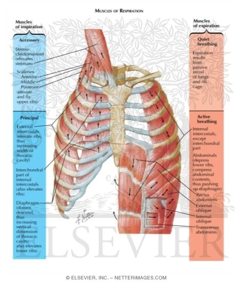
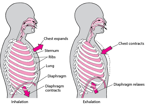
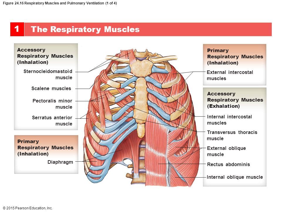
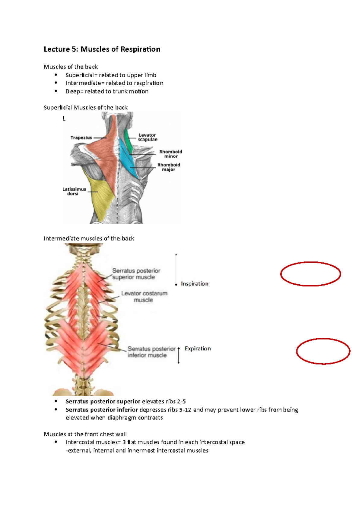
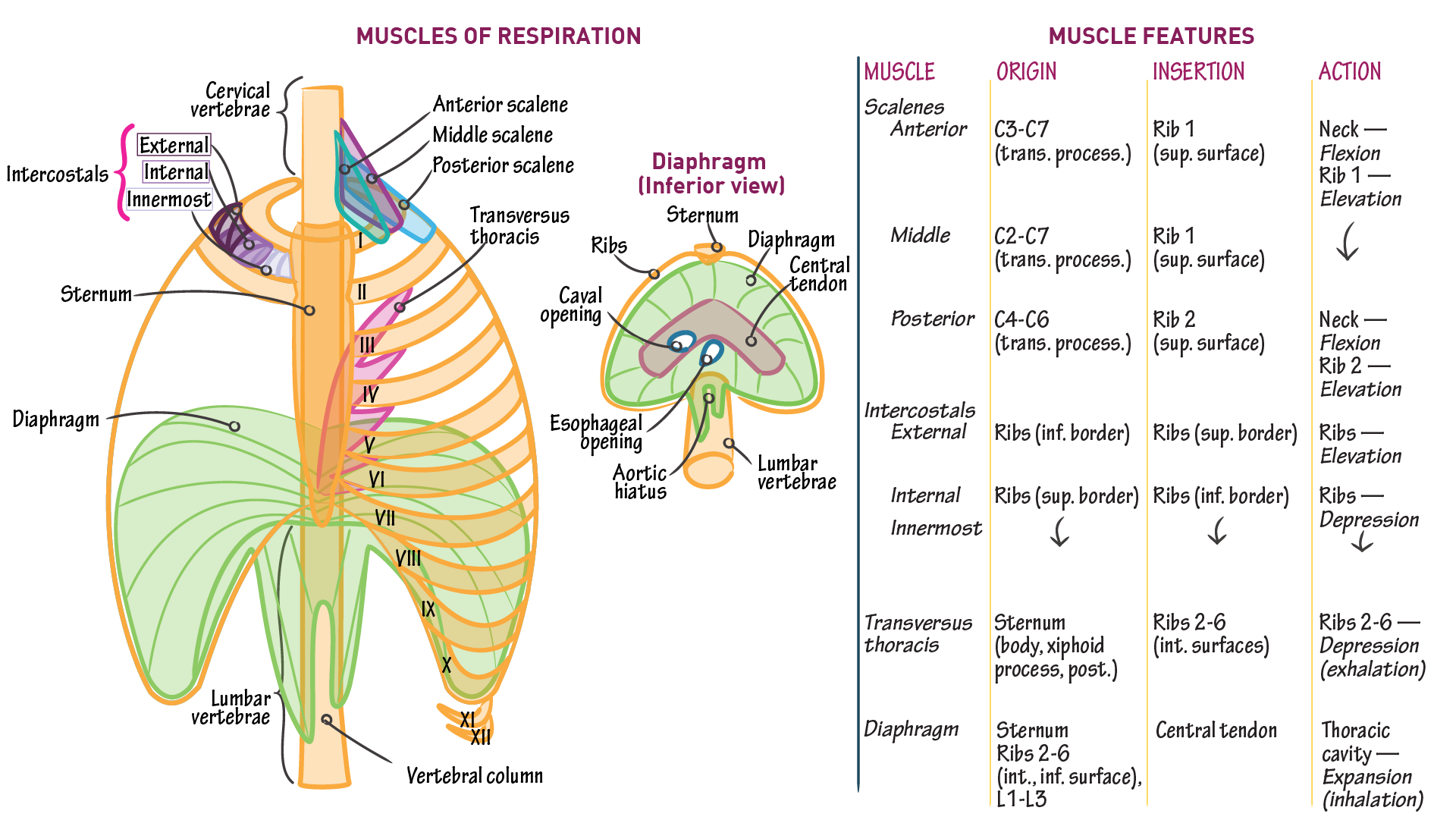
Muscles of respiration - Wikipedia The muscles of respiration are the muscles that contribute to inhalation and exhalation, by aiding in the expansion and contraction of the thoracic cavity.The diaphragm and, to a lesser extent, the intercostal muscles drive respiration during quiet breathing.The elasticity of these muscles is crucial to the health of the respiratory system and to maximize its functional capabilities. Mechanics of Breathing - Inspiration - TeachMePhysiology Inspiration is the phase of ventilation in which air enters the lungs. It is initiated by contraction of the inspiratory muscles: Diaphragm - flattens, extending the superior/inferior dimension of the thoracic cavity. External intercostal muscles - elevates the ribs and sternum, extending the anterior/posterior dimension of the thoracic cavity.
Respiration Control | Boundless Anatomy and Physiology The medulla oblongata is the primary respiratory control center. Its main function is to send signals to the muscles that control respiration to cause breathing to occur. There are two regions in the medulla that control respiration: The ventral respiratory group stimulates expiratory movements. The dorsal respiratory group stimulates ...

Muscles of respiration diagram
Process of Respiration in Humans - Diagram | Healthhype.com Respiration Process. The muscles of respiration contract thereby expanding the chest cavity. This causes a negative pressure within the pleural cavity (where the lungs are housed) which forces the lungs to expand. The expansion of the lungs reduces the air pressure in the lungs. This draws air from the environment which is at a higher pressure. Muscles of Respiration - Physiopedia Action: diaphragm is the main inspiratory muscle, during inspiration it contracts and moves in an inferior direction that increases the vertical diameter of the thoracic cavity and produces lung expansion, in turn, the air is drawn in.[9] Intercostal muscles[edit| edit source] 2.3.3 Muscles for Respiration Diagram | Quizlet Start studying 2.3.3 Muscles for Respiration. Learn vocabulary, terms, and more with flashcards, games, and other study tools.
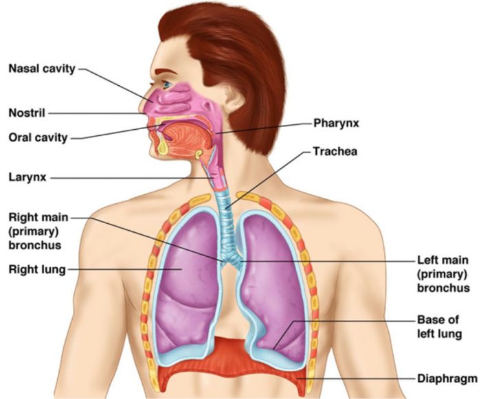
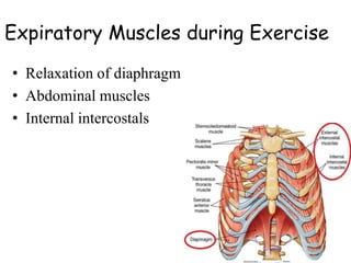
Muscles of respiration diagram. Respiratory System Anatomy, Diagram & Function | Healthline Respiratory. The respiratory system, which includes air passages, pulmonary vessels, the lungs, and breathing muscles, aids the body in the exchange of gases between the air and blood, and between ... The Respiratory System - Diagram, Structure & Function Respiratory system diagram. Lower respiratory tract organs. ... Diaphragm: The diaphragm is a broadband of muscle which sits underneath the lungs, attaching to the lower ribs, sternum and lumbar spine and forming the base of the thoracic cavity. Respiratory system related quizzes. Muscular System - Muscles of the Human Body Attached to the bones of the skeletal system are about 700 named muscles that make up roughly half of a person's body weight. Each of these muscles is a discrete organ constructed of skeletal muscle tissue, blood vessels, tendons, and nerves. Muscle tissue is also found inside of the heart, digestive organs, and blood vessels. Breathing Process in Human Beings (With Diagram ... During forced inspiration and expiration many other muscles besides the diaphragm and intercostal muscles come into play to assist respiration. Most important of this group are the scalene and sternocleidomastoids of the anterior neck muscles which stabilise the first ribs and upper sternum during forced inspiration.
Muscles of Respiration Flashcards | Quizlet these muscles play a role in lifting up upper part of rib cage, have a small role in respiration scalenus muscle anterior, middle, and posterior muscles of the neck that elevate the first and second ribs Muscles of Respiration and How They Work - New Health Advisor Intercostal muscles are one of the most important muscles of respiration and run along the diaphragm. They are attached between the ribs, and allows for changes in the width of the rib cage. Among the 3 layers of the intercostal muscles, the external ones are most important for respiration. Diaphragm: Anatomy, Function, Diagram, Conditions, and ... The diaphragm is the primary muscle used in respiration, which is the process of breathing. This dome-shaped muscle is located just below the lungs and heart. It contracts continually as you ... Muscles of the trunk: Anatomy, diagram, pictures | Kenhub Anterolateral trunk muscles diagram The ... They are also involved in movements of the upper limb and in respiration. Pectoralis major Pectoralis major muscle Musculus pectoralis major 1/4. Pectoralis major is a large, fan-shaped, superficial muscle located on the anterior thoracic wall. It forms the bulk of the chest area and can be easily ...
Respiratory system diagram: Function, facts, conditions ... The diaphragm is a dome-shaped sheet of muscle located below the lungs. It separates the chest from the abdomen. The diaphragm operates as the major muscle of respiration and aids breathing . PDF Cellular Respiration - Exploring Nature Understanding Cellular Respiration Here are three visual depictions of cellular respiration - an equation, an output description and an illustration. 1) Equation: C 6 H 12 O 6 (1 glucose molecule) + 6 O 2 = 6 CO 2 + 6 H 2 O + 36 ATP (ENERGY) carbohydrate + oxygen = carbon dioxide + water + ATP energy 2) Description of the molecules created in all three stages of cellular respiration: muscles of respiration Diagram | Quizlet muscles of respiration Diagram | Quizlet muscles of respiration STUDY Learn Write Test PLAY Match + − Created by Vanessa6873 PLUS Terms in this set (3) internal intercostals ... external intercostals ... diaphragm ... Sets found in the same folder muscles of facial expressions 7 terms Vanessa6873 PLUS muscles of mastification 2 terms Anatomy of breathing: Process and muscles of respiration ... This article will discuss the anatomical basis of breathing and will describe the anatomical components that move every 5 seconds to keep you alive. Contents Thoracic cage Components Ribs Muscles of respiration Thoracic muscles Neck muscles Pectoral girdle muscles Abdominal muscles Airways and lungs Breathing mechanism Inspiration Expiration
Anatomy and Physiology of the Respiratory System Notes ... Chapter 67 Respiratory Physiology: Anatomy & Physiology RESPIRATORY SYSTEM ANATOMY Nose Function: humidifies, warms, filters inspired air; voice resonance chamber; houses olfactory receptors Nasal vibrissae (hairs) coated with mucus → traps large particles (e.g. dust, pollen) Nasal cavity Nasal cavity division Midline nasal septum: composed of septal cartilage, anteriorly Vomer bone ...
Muscles of Respiration: Diagram Diagram | Quizlet Start studying Muscles of Respiration: Diagram. Learn vocabulary, terms, and more with flashcards, games, and other study tools.
2.3.3 Muscles for Respiration Diagram | Quizlet Start studying 2.3.3 Muscles for Respiration. Learn vocabulary, terms, and more with flashcards, games, and other study tools.
Muscles of Respiration - Physiopedia Action: diaphragm is the main inspiratory muscle, during inspiration it contracts and moves in an inferior direction that increases the vertical diameter of the thoracic cavity and produces lung expansion, in turn, the air is drawn in.[9] Intercostal muscles[edit| edit source]
Process of Respiration in Humans - Diagram | Healthhype.com Respiration Process. The muscles of respiration contract thereby expanding the chest cavity. This causes a negative pressure within the pleural cavity (where the lungs are housed) which forces the lungs to expand. The expansion of the lungs reduces the air pressure in the lungs. This draws air from the environment which is at a higher pressure.





:background_color(FFFFFF):format(jpeg)/images/library/12404/anatomy-of-respiratory-system_english.jpg)









0 Response to "39 muscles of respiration diagram"
Post a Comment