38 acl mcl pcl lcl diagram
Download scientific diagram | Concomitant ligament injuries (ACL anterior cruciate liament , MCL medial cruciate liament, PCL posterior cruciate lia- ment) from publication: Lateral ligament ... The ACL crosses in the front of the knee and your Posterior Cruciate Ligament (PCL) crosses along the back of the knee joint. Your Medial Collateral Ligament (MCL) is located on the inner side of your knee. It is a Collateral Ligament that provides stability to the knee. The knee is perhaps the most used major joint in the body, and as such, is ...
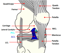
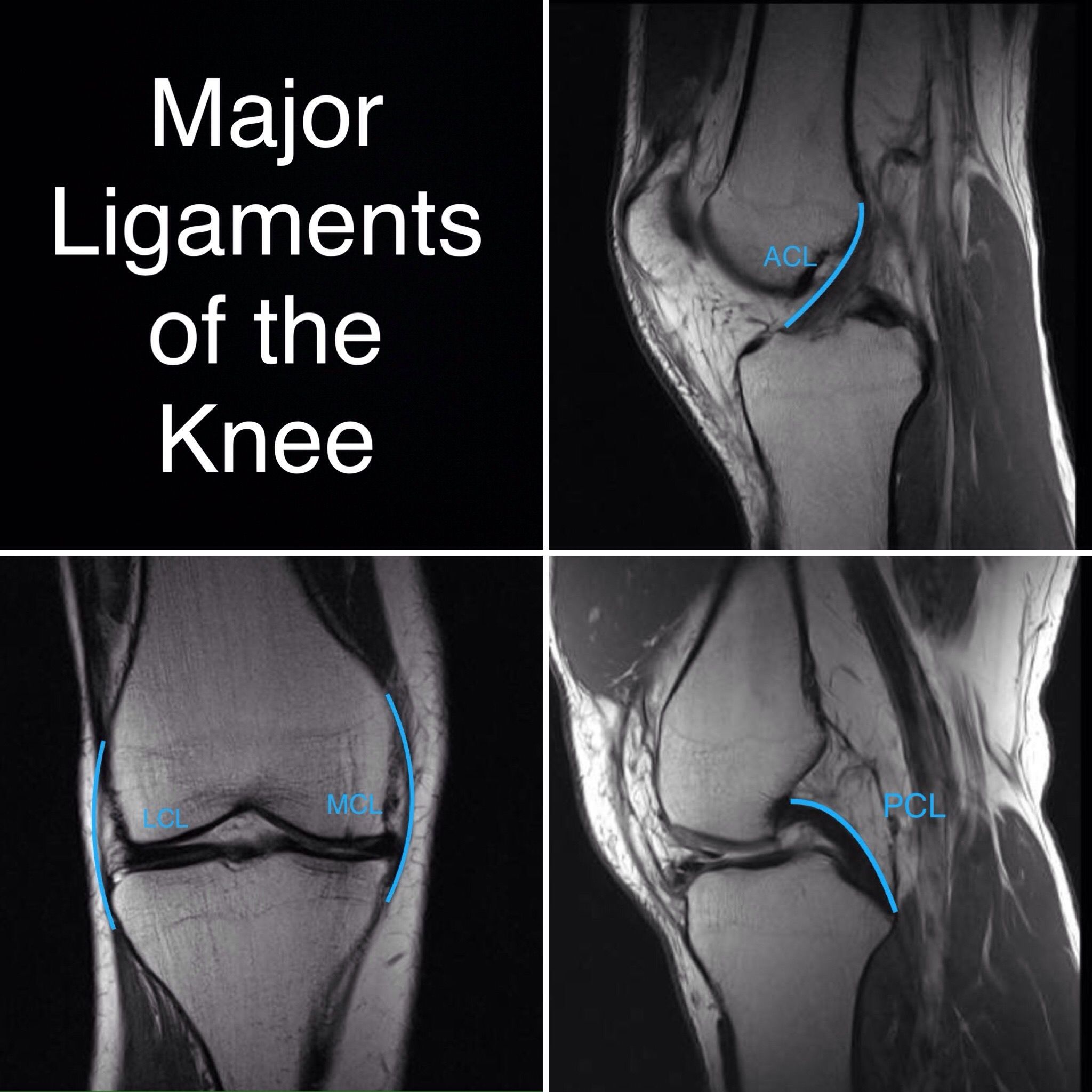
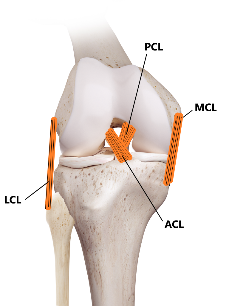
the medial collateral ligament (MCL) (Figure 1a), lateral collateral ligament (LCL) (Figure 1b), anterior cruciate ligament (ACL) (Figure 2a) and posterior cruciate ligament (PCL) (Figure 2b). The MCL connects the femur and tibia medially (on the inside) and resists valgus (knee buckling in) knee motion. A common mechanism of injury

Acl mcl pcl lcl diagram
The medial collateral ligament (MCL) is one of four ligaments that is responsible for keeping the knee joint stable. (The other three are the anterior and posterior cruciate ligaments [ ACL and PCL] and the lateral collateral ligament [ LCL ].) The MCL connects the inner (medial) surfaces of the thigh bone (femur) and the shin bone (tibia) and ... This name is fitting because the ACL crosses the posterior cruciate ligament to form an “X”. It is composed of strong, fibrous material and assists in ... PCL = Posterior Cruciate Ligament (this is the good news, it's OK on me) The stability of the knee largely depends on these four major knee ligaments (the tough, elastic tissues that hold two or more bones together.) The MCL and LCL have sufficient blood flow to heal themselves when torn.
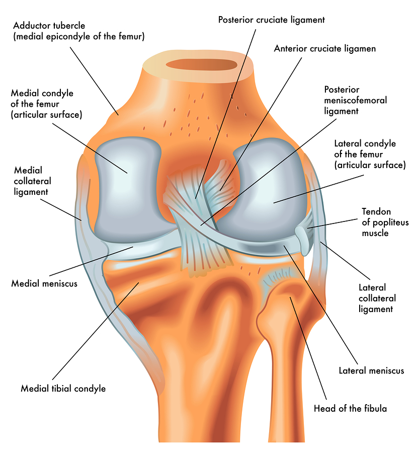
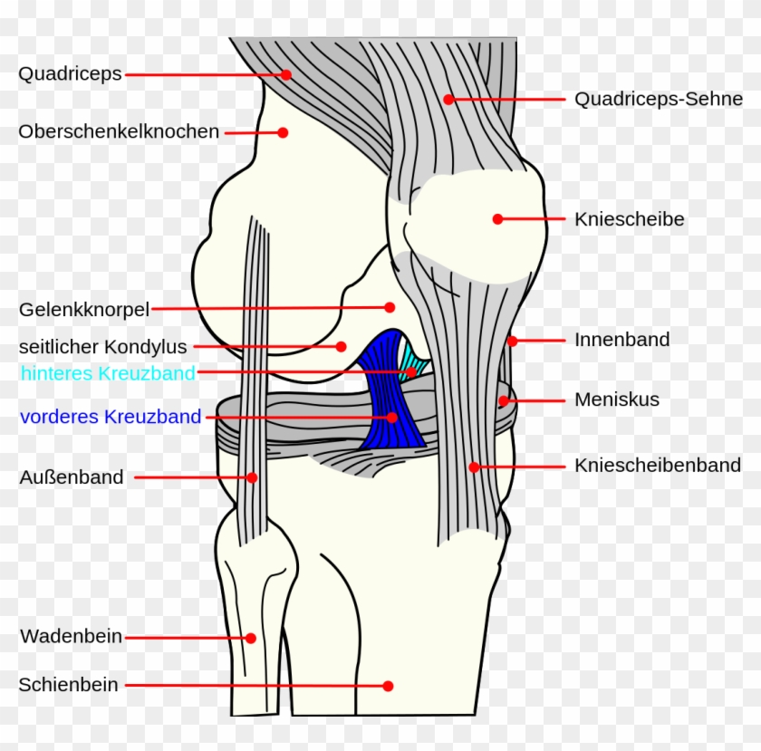
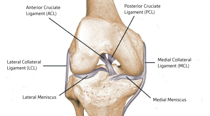
Acl mcl pcl lcl diagram. In the model you can see femur, tibia and fibula, with ACL (anterior cruciate ligament), PCL (posterior cruciate ligament), MCL (medial collateral ligament), LCL (lateral collateral ligament) and medial and lateral meniscus. These are the most typical ligament and cartilage structures in the knee to receive an injury. What is the LCL? Like the ACL and the PCL, the MCL and the lateral collateral ligament (LCL) work together to stabilize the knee joint. The collateral ligaments are located on the outside of the knee and help control the sideways movement of the knee. The LCL is located on the outside of the knee, while the MCL is located on the inside of the ... ACL, MCL, LCL, and PCL All Present One of the four main ligaments of the knee is the anterior cruciate ligament (ACL). Together with the posterior cruciate ligament (PCL), it forms an 'X' shape inside the knee joint. The ACL runs from the bottom of the femur at the back of the knee, diagonally through the joint and attaches to the top of the ... Anterior cruciate ligament (ACL) Posterior cruciate ligament (PCL) Medial Collateral ligament (MCL) Lateral collateral ligament/Posterolateral Corner (LCL/PLC) This specific rehabilitation protocol should be used when both the ACL and PCL are reconstructed, along with one or more of the other ligaments.
A knee ligament injury a sprain of one or more of the four ligaments in the knee, either the Medial Collateral Ligament (MCL), Lateral Collateral Ligament (LCL), Posterior Cruciate Ligament (PCL), or the Anterior Cruciate Ligament (ACL). ACL, PCL, MCL, and LCL injuries are caused by overstretching or tearing of a ligament by twisting or wrenching the knee. Medical Library: Knee - ACL, PCL, MCL, LCL Tear There are four main ligaments in the knee: Anterior Cruciate Ligament, Posterior Cruciate Ligament, Medial Collateral Ligament, and Lateral Collateral Ligament. Tears to any of these ligaments are serious conditions, and may require surgery, or rest and rehabilitation. ACL An anterior cruciate ligament (ACL) tear is an injury to the knee ... By popular demand from yesterday's post [Some Insight on Ekeler's Injury](https://www.reddit.com/r/fantasyfootball/comments/j7ery8/some_insight_on_ekelers_injury/) **Who am I?** By profession I am an Athletic Trainer, I have both my Bachelor's and Master's degree in sports medicine and 10 years of practice in collegiate athletics and Industrial Injury prevention. **Full Disclaimer:** I am not his Doctor, so I do not fully know the extent of his exact injury, but I will shed some light on the... The posterior cruciate ligament (PCL) runs diagonally opposite of the ACL, crossing its path to make an "X" in the middle of the knee. Unlike the ACL, the PCL restricts excessive posterior translation, or sliding backward of the tibia on the femur. This ligament is nearly twice as thick as the ACL, meaning that it is almost twice as strong.
Anterior Cruciate Ligament (ACL).ACL, PCL, MCL, and LCL injuries are caused by overstretching or tearing of a ligament by twisting or wrenching the knee. 31.07.2021 · Segond fracture is an avulsion fracture of the knee that involves the lateral aspect of the tibial plateau and is very frequently (~75% of cases) associated with disruption of the anterior cruciate ligament (ACL).On the frontal ... the Posterior Cruciate Ligament (PCL) and Anterior Cruciate Ligament (ACL) control back-and-forth movement; the Medial Collateral Ligament (MLC) helps to brace the inside of the knee; the Lateral Collateral Ligament (LCL) braces the ouside of the knee, controlling sideways motion and protecting the knee from over-extending. While most injuries ... ACL vs. MCL tear. The ACL is located inside of the knee joint and connects the top front of the tibia (shinbone) to the bottom back of the femur (thighbone). Together, the ACL and PCL cross each other to form an "X" inside of your knee. The ACL prevents the tibia from sliding too far forward relative to the femur and is important for ... Ligament Injury (ACL, PCL, MCL, LCL & multiligament injury) ... Diagram of Anterior Cruciate Ligament - Knee Surgeon Melbourne ...
MCL Tear Symptoms. Just as with an ACL tear, you may hear a popping sound when the injury occurs. Some of the most common symptoms of an MCL tear include: Pain on the inner side of the knee. Feeling the knee "give out". Swelling. The knee feels unstable. Pain when putting weight on the knee. Locked knee.
These are the four main ligaments or tough bands of tissue in the knee. ACL is the anterior cruciate ligament, which connects the thigh bone to the shin bone.
Different Types Of Knee Injuries - ACL, LCL, MCL and PCL. Our knees are one of the most vital joints in our bodies, shouldering our weight and facilitating movements like walking, running and sitting. Your knee is made up of four distinct ligaments, with one on the front, back and each side. Damage to the knee can result in a partial or ...
We are pleased to provide you with the picture named Pcl Acl Lcl Mcl Meniscus Anatomy.We hope this picture Pcl Acl Lcl Mcl Meniscus Anatomy can help you study and research. for more anatomy content please follow us and visit our website: www.anatomynote.com. Anatomynote.com found Pcl Acl Lcl Mcl Meniscus Anatomy from plenty of anatomical pictures on the internet.
Anterior cruciate ligament (ACL) is the most commonly injured knee ligament.It connects the thigh bone to the shin bone. Posterior cruciate ligament (PCL) also links the thigh bone to the shin ...
Posterior cruciate ligament or PCL - located at the back of the knee, controls backward movement of the shin bone; Medial collateral ligament or MCL - connects the thigh bone to the shin bone on the inside of the knee—MCL stabilizes the inner knee
Anterior Cruciate Ligament Repair (ACL, MCL, MPFL, PCL, LCL) The ACL, or anterior cruciate ligament, is one of the ligaments in the knee. Athletes who play sports that require sudden stops and turns are more likely to experience injuries or tears to their ACLs, and tears usually have to be reconstructed surgically.
Associate Professor of Orthopaedics Chief - Division of Sports Medicine Tel: (212) 598-6784 Rehabilitation Protocol: ACL, MCL and PCL Reconstruction
Surgery (ACL, PCL, MCL) Depending on the severity and type of ligament injury, surgery may be recommended. For ACL injuries, arthroscopic or open surgery is done using a graft to replace the damaged ligament. For certain PCL cases where the ligament is no longer attached properly to the shinbone, surgery is considered.
Acl And Mcl Diagram. Signs and symptoms of a medial collateral ligament (MCL) injury include The anterior cruciate ligament (ACL) and posterior cruciate ligament. The MCL is one of four major ligaments that supports the knee. either partial or complete ruptures in the ligament significantly increases the load on the ACL.
An LCL injury can be a stretch, partial tear, or a complete tear of the ligament. The injury usually occurs when the knee joint is pushed from the inside, which results in stress being placed on the outside part of the joint. An LCL injury often occurs along with other ligament injuries, including ACL, PCL, and MCL, and is frequently seen along ...
Treatment. Treatment for ACL and PCL injuries essentially is the same, but will differ depending on the severity, or grade, of the injury: Grade 1: The ligament is slightly stretched but the knee is stable. Grade 2: The ligament has become loose or is partially torn. Grade 3: There is a complete rupture of the ligament.
ACL vs. MCL tears: Although symptoms of ACL and MCL tears are similar, a few key differences will help identify whether the injury affected the ACL or MCL. An ACL tear will have a more distinctive and loud popping sound than an MCL tear. The location of your pain and swelling could indicate either an ACL or MCL tear.
b a FIGURE 2.1: (a) Schematic representation of the knee ligaments (ACL, PCL, MCL and LCL) as 1D, 2D and 3D elements adopted from (Galbusera et al., 2014 ) (b) ...
Pcl posterior cruciate ligament strain or tear. The 2 ligaments are also called cruciform ligaments as they are arranged in a crossed formation. When the knee is fully extended both cruciate ligaments are taut and the knee is locked. There is both an anterior cruciate ligament acl and a posterior cruciate ligament pcl.
The posterior cruciate ligament (PCL) is about two inches long and connects the femur to the tibia at the back of the knee. It limits the backward or posterior motion of the tibia (shinbone). Twisting or overextending the knee can cause the PCL to tear, leaving the knee unstable and potentially unable to support a person's full body weight.
Jun 24, 2021 — MEDICALLY REVIEWED Knee ligaments are bands of tissue that connect your thigh bone to your lower leg bones. They help stabilize your knee ...
Hi everyone, This week there was a thrilling Merseyside derby with high levels of play between both teams but also some really worrying situations with dodgy decisions from refs and reckless challenges. The most concerning being the Jordan Pickford challenge on Virgil Van Dijk. How this didn’t get a red card is an utter joke but I’m also hearing VAR didn’t check it which makes the situation even worse in my opinion. If you don’t know me already, my name is Matthew Feyissa and I am a medical st...
PCL = Posterior Cruciate Ligament (this is the good news, it's OK on me) The stability of the knee largely depends on these four major knee ligaments (the tough, elastic tissues that hold two or more bones together.) The MCL and LCL have sufficient blood flow to heal themselves when torn.
This name is fitting because the ACL crosses the posterior cruciate ligament to form an “X”. It is composed of strong, fibrous material and assists in ...
The medial collateral ligament (MCL) is one of four ligaments that is responsible for keeping the knee joint stable. (The other three are the anterior and posterior cruciate ligaments [ ACL and PCL] and the lateral collateral ligament [ LCL ].) The MCL connects the inner (medial) surfaces of the thigh bone (femur) and the shin bone (tibia) and ...

















0 Response to "38 acl mcl pcl lcl diagram"
Post a Comment