38 gram positive bacteria diagram
Flow Chart Of The Study Design Each Patient Having A First Positive Scientific Diagram. Flowchart For Identification Of Anaerobic Gram Positive Bacilli 1 Scientific Diagram. Gram Negative Bacteria Positive Microbiology Flowchart Others Angle Text Infection Png Pngwing. Starting With Figure 17 5 And Using From Case Study T Chegg. Download scientific diagram | listeria monocytogenes gram stain as seen in a light microscope (kenneth, 2004). Listeria is a kind of gram positive rod bacteria, with the size of. Motility test medium (mtm, difco) (m103) . Studies on the isolation of . The pathogenic bacterium listeria monocytogenes can persist in food.
4 Bacteria: Cell Walls . It is important to note that not all bacteria have a cell wall.Having said that though, it is also important to note that most bacteria (about 90%) have a cell wall and they typically have one of two types: a gram positive cell wall or a gram negative cell wall.. The two different cell wall types can be identified in the lab by a differential stain known as the Gram stain.
Gram positive bacteria diagram
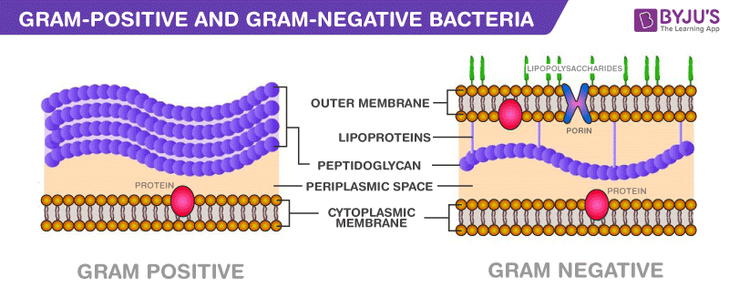
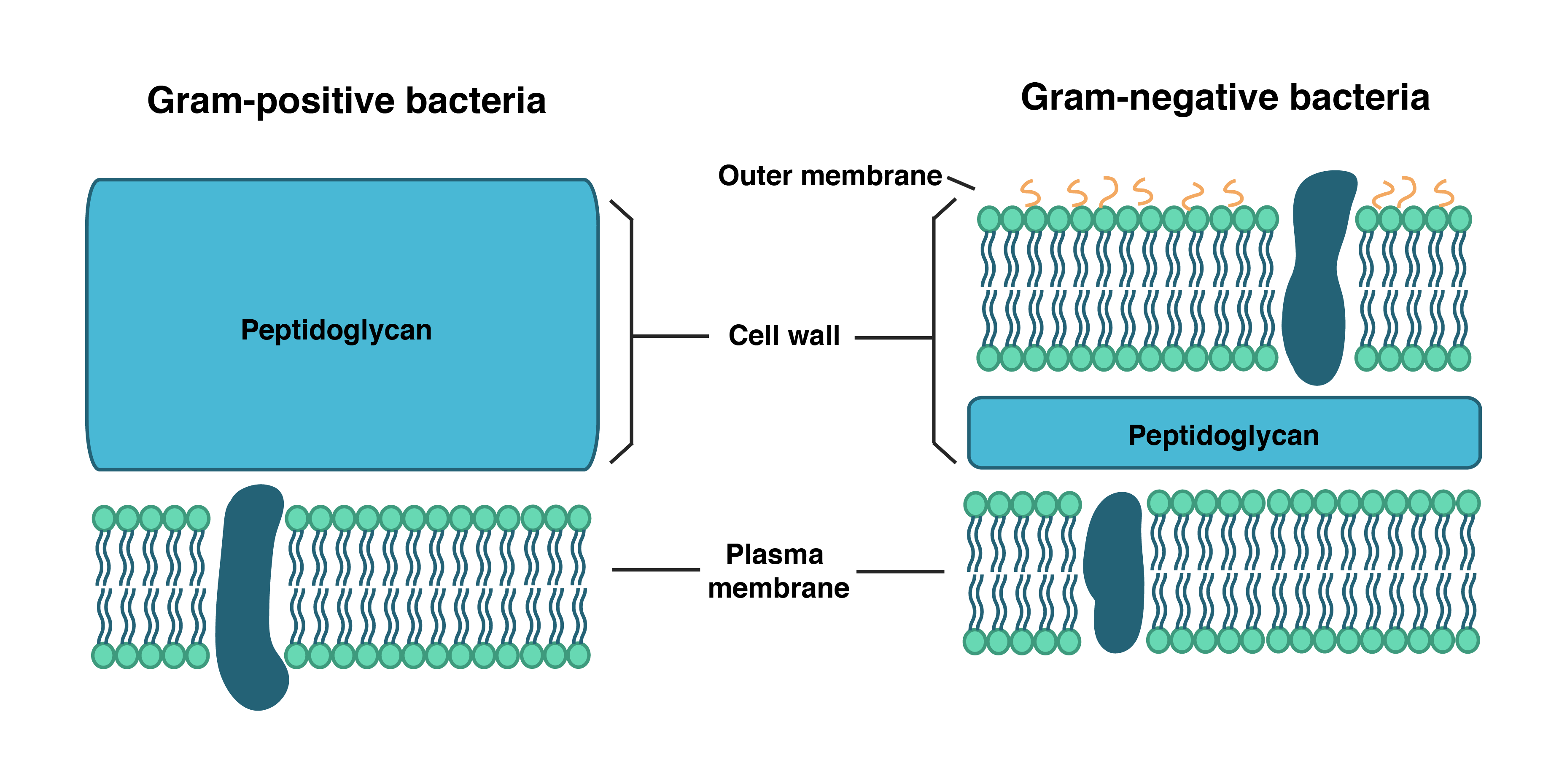
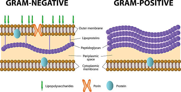
The Gram stain is a differential staining technique used to classify & categorize bacteria into two major groups: Gram positive and Gram negative, based on the differences of the chemical and physical properties of the cell wall. In Gram-positive cells, peptidoglycan makes up as much as 90% of the thick cell wall; more than 20 layers of peptidoglycan stacked together. These layers are the outermost cell wall structure of Gram+ cells, whereas in Gram-negative cells, the thinner peptidoglycan component is covered by an external lipopolysaccharide (LPS) membrane. Gram-positive bacteria, on the other hand, retains the gram stain and show a visible violet colour upon the application of mordant (iodine) and ethanol (alcohol). Gram-positive bacteria constitute a cell wall, which is mainly composed of multiple layers of peptidoglycan that forms a rigid and thick structure.
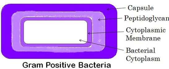
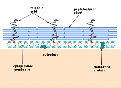
Gram positive bacteria diagram. Start studying Microbiology: Compare and contrast gram-positive and gram-negative bacteria. Learn vocabulary, terms, and more with flashcards, games, and other study tools. Gram positive bacteria are a group of organisms that fall under the phylum Firmicutes (however, a few species have a Gram negative cell wall structure). As compared to Gram negative bacteria, this group of bacteria is characterized by their ability to retain the primary stain (Crystal violet) during Gram staining (giving a positive result). ... The walls of gram-positive bacteria have simpler chemical structures compared to gram-negative bacteria. Gram-positive cell wall. Gram-positive cell wall is thick measuring about 15-80 nm and more homogenous compared to gram-negative cell wall. This cell wall consists of large amount of peptidoglycan arranged in several layers. Gram-positive bacteria comprise cocci, bacilli, or branching filaments. Health professionals need to understand the important difference between gram-positive and gram-negative bacteria. Gram-positive bacteria are bacteria classified by the color they turn in the staining method. Hans Christian Gram developed the staining method in 1884.
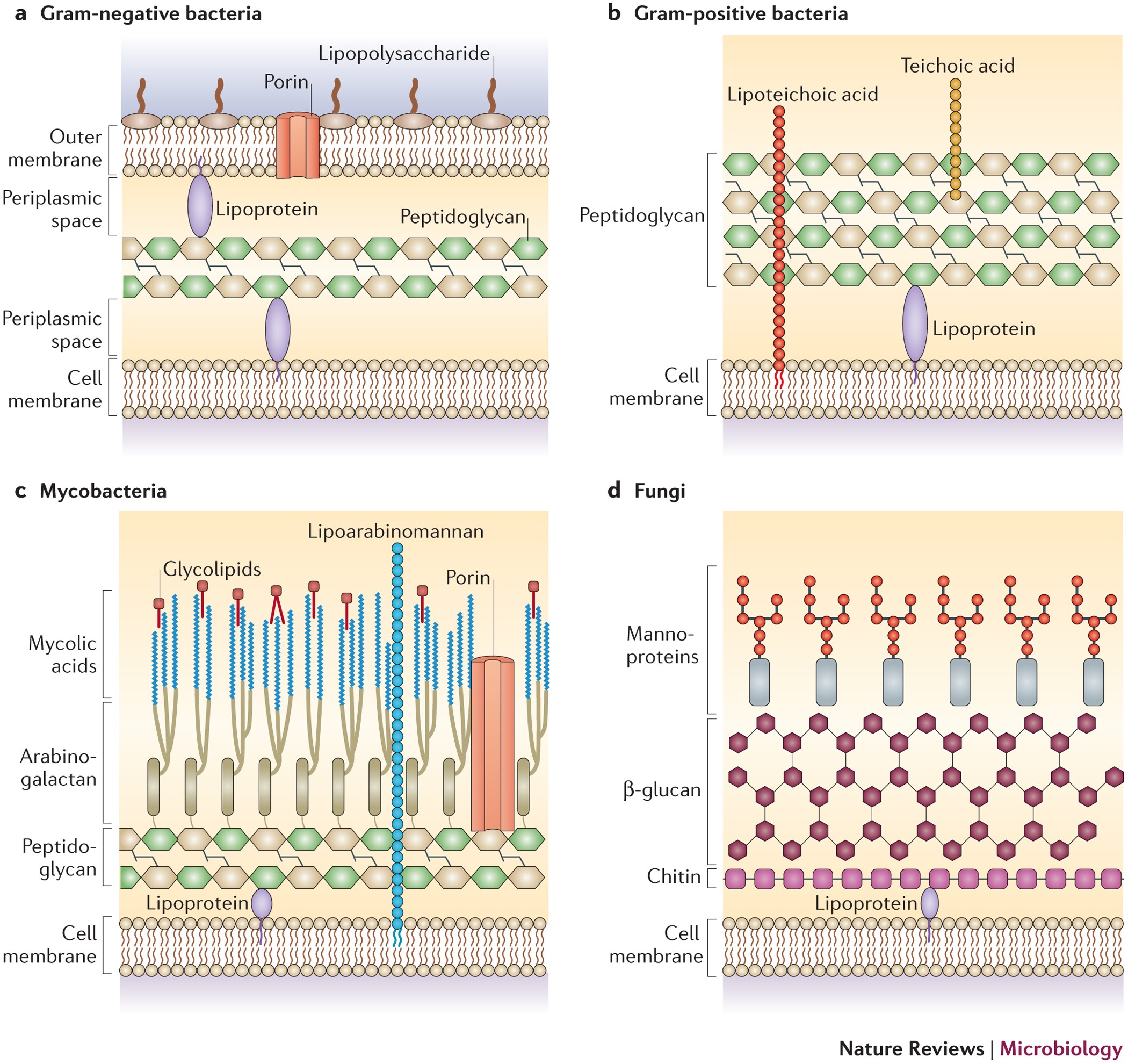
S. pyogenes is a gram-positive bacterium. Gram-positive bacteria have structural differences that distinguish them from gram-negative organisms. Of particular interest to this case is the fact that these differences in structure can be exploited during treatment. Drag each of the labels onto the diagram to correctly label the indicated structures. Compare and contrast the cell walls of typical Gram-positive and Gram-negative bacteria. 3. Relate bacterial cell wall structure to the Gram-staining reaction. 37 . 38 Bacterial Cell Wall • Peptidoglycan (murein) -rigid structure that lies just outside the cell plasma membrane Best Gram Positive Bacteria Stock Photos, Pictures from media.istockphoto.com Been used as a marker to distinguish listeria monocytogenes from other. Download scientific diagram | listeria monocytogenes gram stain as seen in a light microscope (kenneth, 2004). For figure 1, please refer to the contributing authors page for the . Bacteria can be divided into two major groups: Gram positive and Gram negative, based on the Gram stain reaction. Gram-positive organisms have a thick cell wall, together with teichoic acids. Gram-negative organisms have a thin cell wall and an outer envelope containing lipopolysaccharides and lipoproteins.
Both Gram-positive and Gram-negative bacteria take up the same amounts of crystal violet (CV) and iodine (I). The CV-I complex, however, is trapped inside the Gram-positive cell by the dehydration and reduced porosity of the thick cell wall as a result of the differential washing step with 95 percent ethanol or other solvent mixture. The diagram below illustrates the differences in the structure of Gram positive and Gram negative bacteria. The two key features that lead to the differing visualization properties of Gram positive and Gram negative species are the thickness of the peptidoglycan layer and presence or absence of the outer lipid membrane. For the gram-positive cell wall, it has a thickness of about 20-80nm thickness made up of a thick peptidoglycan layer outside its cell membrane, unlike the thin layer of gram-negative bacteria (10-15nm) which has a very thin layer of the peptidoglycan of 2-7nm but has a thicker lipid layer making it quite complex than the Gram-positive cell wall. Gram-positive bacteria are those that are stained dark blue or violet by Gram staining.This is in contrast to gram-negative bacteria, which cannot hold the crystal violet stain. Instead they take up the counterstain (safranin or fuchsine) and appear red or pink.The difference is caused by the cell wall structure. Gram-positive organisms have thick peptidoglycan layer.
1
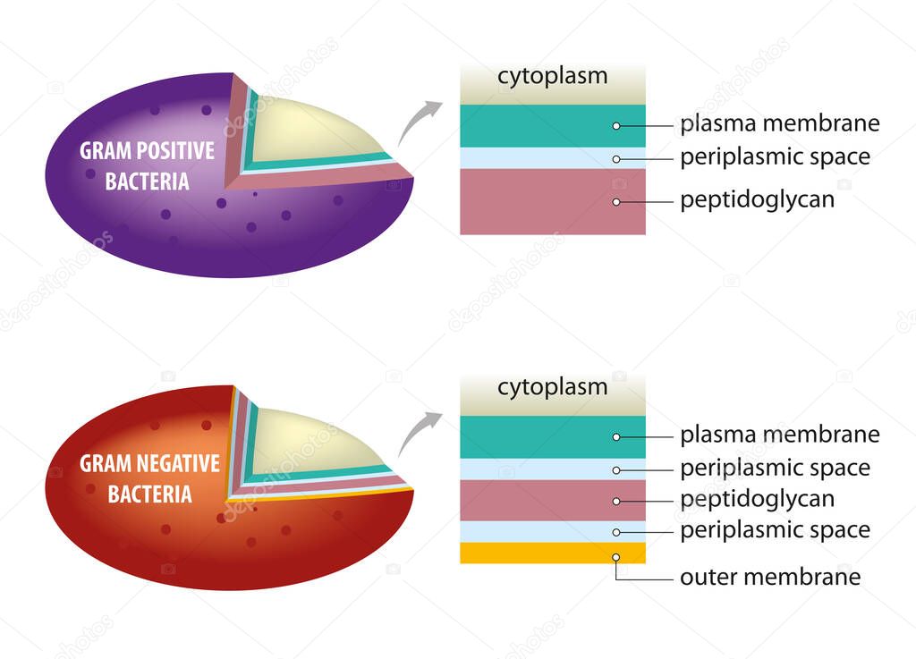
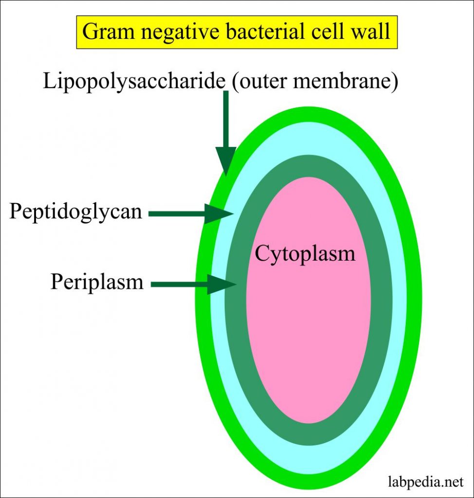
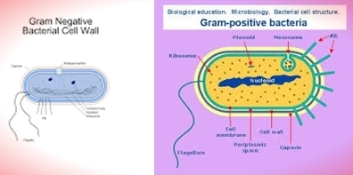
The cell wall structure of Gram negative bacteria is more complex than that of Gram positive bacteria. Located between the plasma membrane and the thin peptidoglycan layer is a gel-like matrix called periplasmic space. Unlike in Gram positive bacteria, Gram negative bacteria have an outer membrane layer that is external to the peptidoglycan ...

Gram Positive Bacterial Cell Envelopes The Impact On The Activity Of Antimicrobial Peptides Sciencedirect
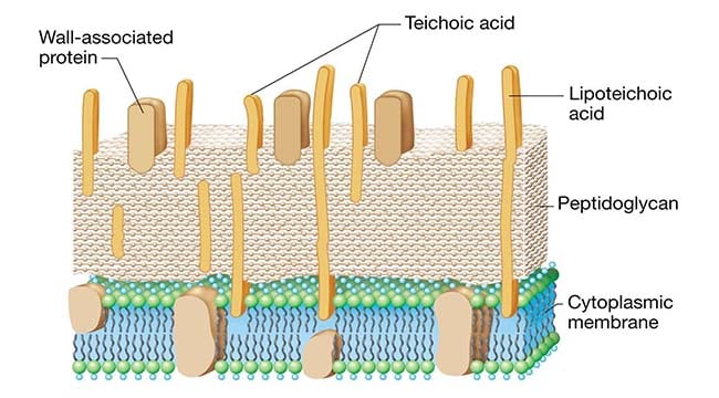
Differences between Gram-positive and Gram-negative bacteria cell wall The gram-Positive Cell wall of Bacteria. Bacterial cell wall that is gram-positive contains peptidoglycan and teichoic acids with some species having additional carbohydrates and proteins.The murein component is what gives shape to the gram-positive bacterial cell wall; it also helps the bacteria cells to resist osmotic ...

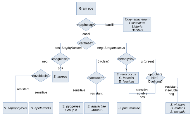
Gram Positive Cocci Bacteria Identification Algorithm Catalase Grepmed
4,312 gram positive stock photos, vectors, and illustrations are available royalty-free. See gram positive stock video clips. of 44. gram positive rod gram positive cell gram positive and gram negative gram positive bacteria bacteria cell wall streptococus pneumoniae germ layers wall gram bacteria sample gram positive and negative bacteria.

Gram Positive Vs Gram Negative Technology Networks
Prokaryotes are identified as gram-positive if they have a multiple layer matrix of peptidoglycan forming the cell wall. Crystal violet, the primary stain of the Gram stain procedure, is readily retained and stabilized within this matrix, causing gram-positive prokaryotes to appear purple under a brightfield microscope after Gram staining. For many years, the retention of Gram stain was one of ...
Gram Positive Bacteria Biology Ease
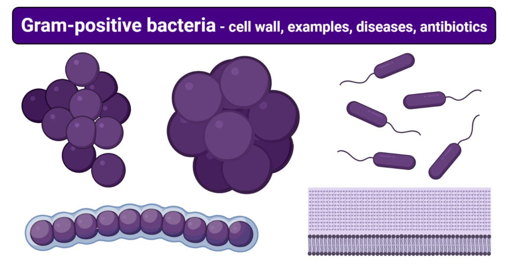
Gram-positive bacteria are the genus of bacteria family and a member of the phylum Firmicutes. These bacteria retain the colour of the crystal violet stain which is used during gram staining. These bacteria give a positive result in the Gram stain test by appearing purple coloured when examined under a microscope, hence named, gram-positive ...
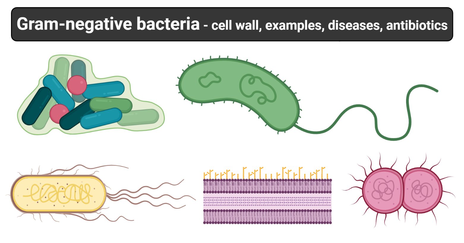
Gram Negative Bacteria Cell Wall Examples Diseases Antibiotics
Gram positive bacteria have a large peptidoglycan structure. As noted above, this accounts for the differential staining with Gram stain. Some Gram positive bacteria are also capable of forming spores under stressful environmental conditions such as when there is limited availability of carbon and nitrogen. Spores therefore allow bacteria to ...
Major Difference Between Gram Positive And Gram Negative Bacteria
Gram Staining is the common, important, and most used differential staining techniques in microbiology, which was introduced by Danish Bacteriologist Hans Christian Gram in 1884. This test differentiate the bacteria into Gram Positive and Gram Negative Bacteria, which helps in the classification and differentiations of microorganisms.
Schematic Illustration Of Difference Between Bacterial Cell Wall Gram Positive And Gram Negative Premium Vector In Adobe Illustrator Ai Ai Format Encapsulated Postscript Eps Eps Format
In bacteriology, gram-positive bacteria are bacteria that give a positive result in the Gram stain test, which is traditionally used to quickly classify bacteria into two broad categories according to their type of cell wall.. Gram-positive bacteria take up the crystal violet stain used in the test, and then appear to be purple-coloured when seen through an optical microscope.
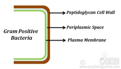
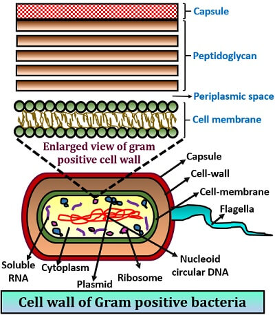
Cell Wall Of Gram Positive Bacteria Easybiologyclass Easy Biology Class
In Gram-positive bacteria, peptidoglycan makes up as much as 90% of the thick cell wall enclosing the plasma membrane. Gram Staining! During Gram staining, these thick, multiple layers (20-80 nm) of peptidoglycan retain the dark purple primary stain crystal violet, whereas Gram-negative bacteria stain pink.
Structural Comparison Of Gram Image Eurekalert Science News Releases
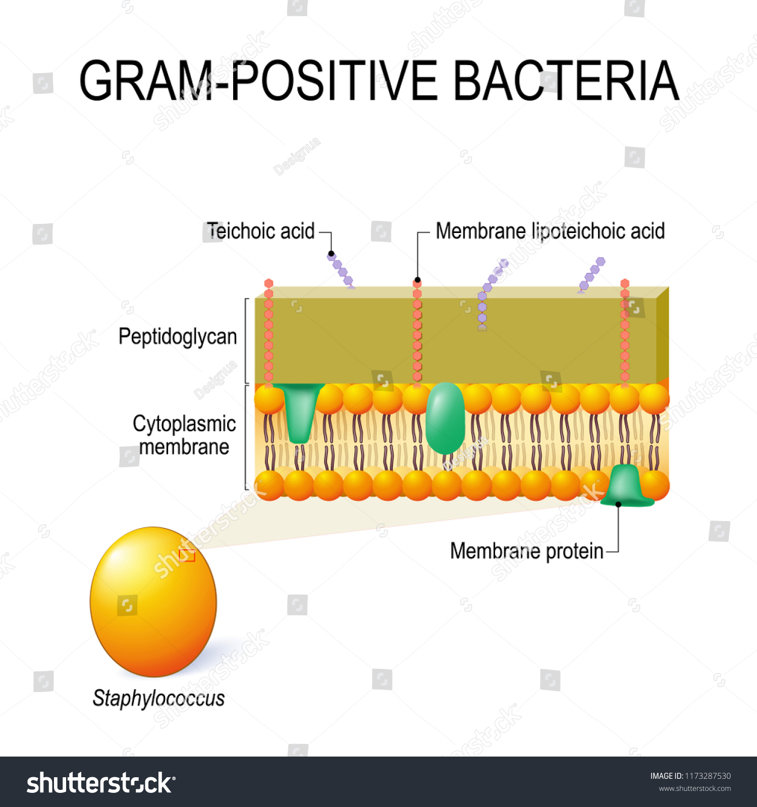
8. According to Peberdy (1980) the only compound present in the cell walls of both Gram-negative and Gram-positive bacteria is 'peptidoglycan'. The cell walls of Gram-positive bacteria contain up to 95% peptidoglycan and up to 10% teichoic acids. 9. Cytoplasmic membrane is a thin (5-10 nm) layer lining the inner surface of the cell wall.
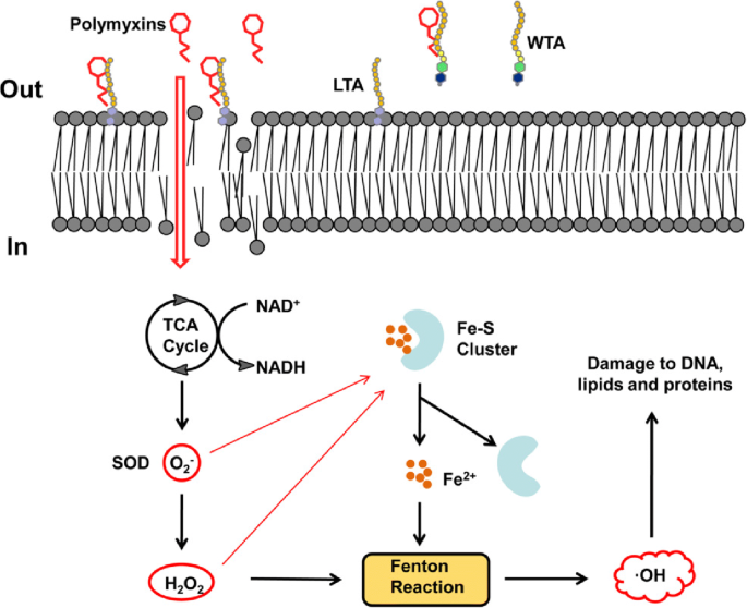
Mechanisms Of Bactericidal Action And Resistance Of Polymyxins For Gram Positive Bacteria Springerlink
Pathogenic bacteria are bacteria that can cause disease. This article focuses on the bacteria that are pathogenic to humans. Most species of bacteria are harmless and are often beneficial but others can cause infectious diseases.The number of these pathogenic species in humans is estimated to be fewer than a hundred. By contrast, several thousand species are part of the gut flora present in ...

Cell Wall Structures Of Gram Positive And Gram Negative Bacteria And Download Scientific Diagram
Gram-positive bacteria, on the other hand, retains the gram stain and show a visible violet colour upon the application of mordant (iodine) and ethanol (alcohol). Gram-positive bacteria constitute a cell wall, which is mainly composed of multiple layers of peptidoglycan that forms a rigid and thick structure.
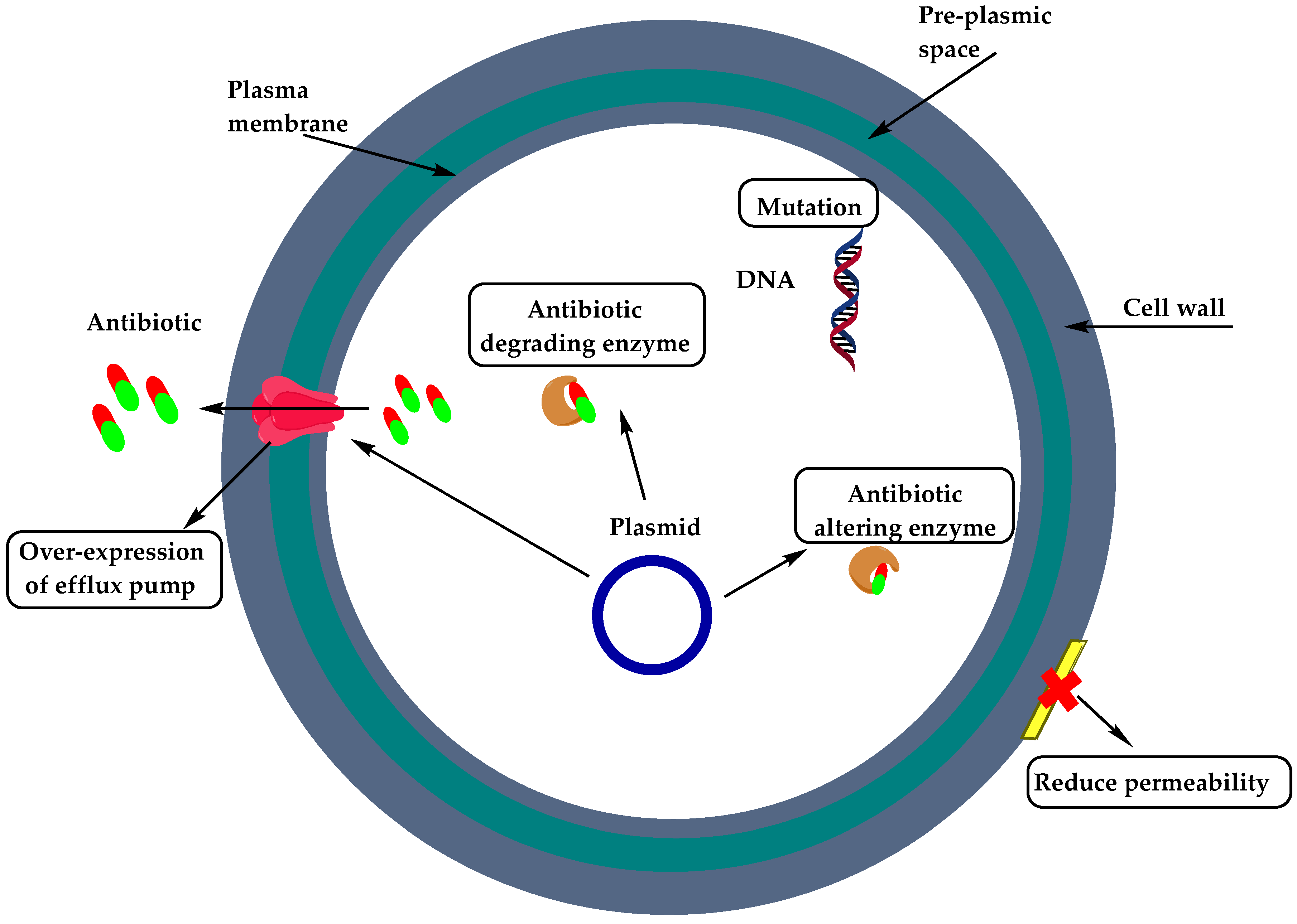
Molecules Free Full Text Resistance Of Gram Positive Bacteria To Current Antibacterial Agents And Overcoming Approaches
In Gram-positive cells, peptidoglycan makes up as much as 90% of the thick cell wall; more than 20 layers of peptidoglycan stacked together. These layers are the outermost cell wall structure of Gram+ cells, whereas in Gram-negative cells, the thinner peptidoglycan component is covered by an external lipopolysaccharide (LPS) membrane.
:max_bytes(150000):strip_icc()/gram_negative_bacteria-5b7f308646e0fb002cbcdc5d.jpg)
Gram Positive Vs Gram Negative Bacteria
The Gram stain is a differential staining technique used to classify & categorize bacteria into two major groups: Gram positive and Gram negative, based on the differences of the chemical and physical properties of the cell wall.

Gram Positive Bacteria Gram Negative Bacteria Gram Stain Coccus Classification Angle Text Png Pngegg
Bacterial Cell Wall Structure Composition And Types Online Biology Notes

Get Portable Version For Macbook Yosemite Gram Positive And Negative Bacteria 1 0 0019 Via Isohunt Nwb Sln My First Jugem
Difference Between Gram Positive And Gram Negative Bacteria With Comparison Chart Video Biology Reader
Through The Wall Extracellular Vesicles In Gram Positive Bacteria Mycobacteria And Fungi Nature Reviews Microbiology
Vektor Stok Cell Wall Structure Grampositive Bacteria Example Tanpa Royalti 1173287530

Endotoxin Gram Negative Bacteria Gram Positive Bacteria Bacterial Cell Structure Cell Wall Bacteria Angle Text Png Pngegg

Protein Secretion And Surface Display In Gram Positive Bacteria Philosophical Transactions Of The Royal Society B Biological Sciences
Gram Stain Gram Stain Procedure Labpedia Net

Gram Positive Bacteria Microbiology Medbullets Step 1
1
Difference Between Gram Positive Bacteria And Gram Negative Bacteria
740 Gram Positive Bacteria Stock Photos Pictures Royalty Free Images Istock
Gram Positive Bacteria Wikipedia
Gram Positive Cell Wall Microbe Notes
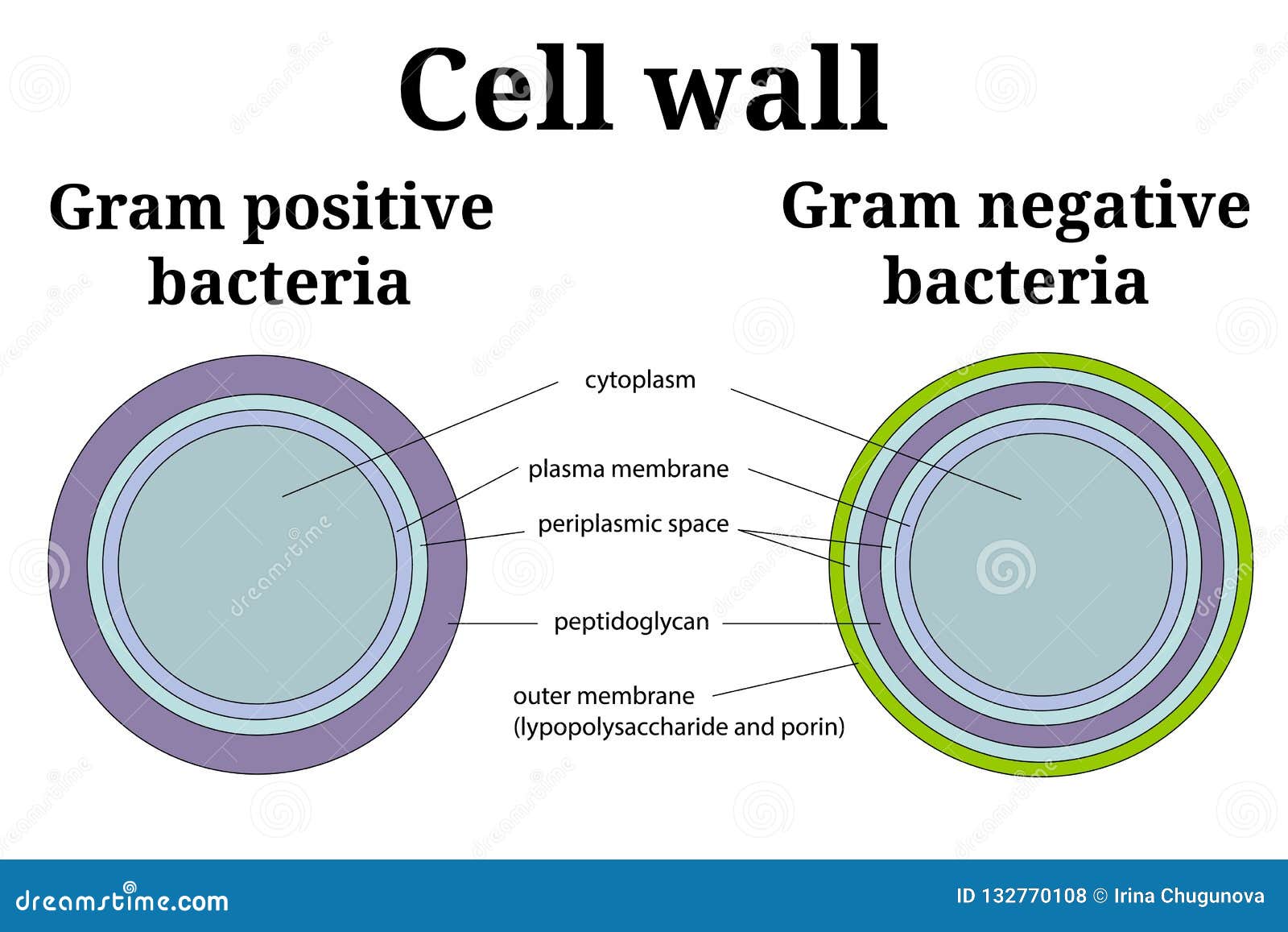
Bacteria Cell Wall Illustration Gram Positive And Gram Negative Cell Wall Differents Stock Vector Illustration Of Cytoplasm Medicine 132770108

Gram Positive Vs Gram Negative Bacteria Microbe Online
Structure And Function Of Bacterial Cells

Gram Positive Vs Gram Negative Technology Networks

Difference Bacteria Cell Walls Of Gram Positive And Gram Negative Download Scientific Diagram

Quia 9ap Chapter 27 Bacteria And Archaea Detailed Cell Wall Plasma Membrane Microbiology
Gram Positive Bacteria Cell Wall Examples Diseases Antibiotics

Bacteria Cell Wall Gram Positive And Gram Negative

Simplified Scheme Of Qs Network In Gram Positive Bacteria Qs In Gram Positive Bacteria Transparent Png 850x538 Free Download On Nicepng
0 Response to "38 gram positive bacteria diagram"
Post a Comment