38 labeled diagram of a microscope
Microscope, Microscope Parts, Labeled Diagram, and Functions Microscope, Microscope Parts, Labeled Diagram, and Functions Published by Admin on January 19, 2022 January 19, 2022. What is Microscope? A microscope is a laboratory instrument used to examine objects that are too small to be seen by the naked eye. It is derived from Ancient Greek words and composed of mikrós, "small" and skopeîn,"to ... PDF Parts of a Microscope Printables - Homeschool Creations Label the parts of the microscope. You can use the word bank below to fill in the blanks or cut and paste the words at the bottom. Microscope Created by Jolanthe @ HomeschoolCreations.net eyepiece head objective lenses arm focusing knob base illuminator stage stage clips nosepiece.
A Study of the Microscope and its Functions With a Labeled ... A Study of the Microscope and its Functions With a Labeled Diagram To better understand the structure and function of a microscope, we need to take a look at the labeled microscope diagrams of the compound and electron microscope. These diagrams clearly explain the functioning of the microscopes along with their respective parts.

Labeled diagram of a microscope
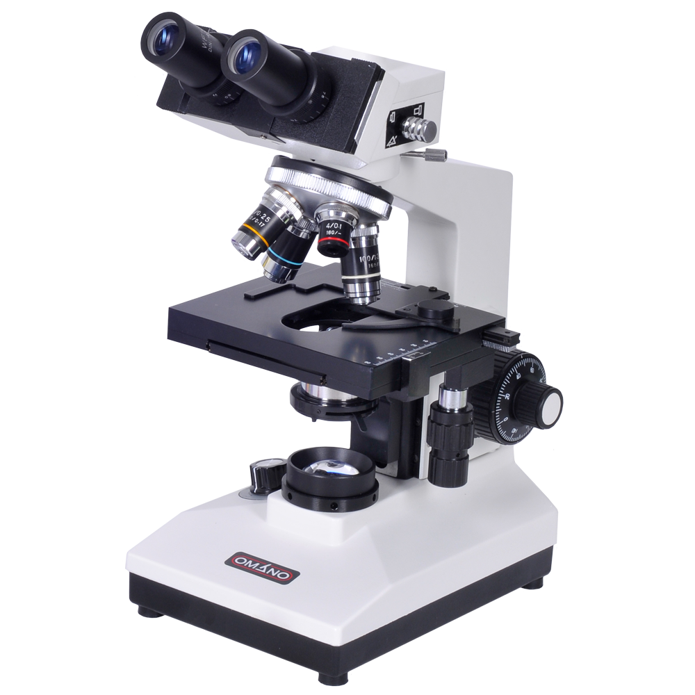
Light Microscope- Definition, Principle, Types, Parts ... Figure: Labeled Diagram of a Light Microscope. Types of light microscopes (optical microscope) With the evolved field of Microbiology, the microscopes. used to view specimens are both simple and compound light microscopes, all using lenses. The difference is simple light microscopes use a single lens for magnification while compound lenses use ... Compound Microscope Parts, Functions, and Labeled Diagram ... So, a compound microscope with a 10x eyepiece magnification looking through the 40x objective lens has a total magnification of 400x (10 x 40). Specimen or slide: The object used to hold the specimen in place along with slide covers for viewing. ... Compound Microscope Parts, Functions, and Labeled Diagram. Parts of a Compound Microscope. Labelled Diagram of Compound Microscope - Biology Discussion The below mentioned article provides a labelled diagram of compound microscope. Part # 1. The Stand: The stand is made up of a heavy foot which carries a curved inclinable limb or arm bearing the body tube. The foot is generally horse shoe-shaped structure (Fig. 2) which rests on table top or any other surface on which the microscope in kept.
Labeled diagram of a microscope. Labeling the Parts of the Microscope | Microscope World ... Labeling the Parts of the Microscope. This activity has been designed for use in homes and schools. Each microscope layout (both blank and the version with answers) are available as PDF downloads. You can view a more in-depth review of each part of the microscope here. Animal Cell Diagram Under Light Microscope Labeled ... Tuesday, April 20th 2021. | Diagram. Animal Cell Diagram Under Light Microscope. To make observations and draw scale. This shows a generalized animal cell under a light microscope. We all keep in mind that the human physique is amazingly elaborate and one way I discovered to comprehend it is by way of the style of human anatomy diagrams. Microscope Parts and Functions With Labeled Diagram and ... Microscope Parts and Functions With Labeled Diagram and Functions How does a Compound Microscope Work?. Before exploring microscope parts and functions, you should probably understand that the compound light microscope is more complicated than just a microscope with more than one lens.. First, the purpose of a microscope is to magnify a small object or to magnify the fine details of a larger ... Plant Cell Under Light Microscope Labeled - Diagram Sketch Plant Cell Under Light Microscope Labeled. angelo on October 4, 2021. Editible Eps Vector File The Animal Cell Diagram Vector Etsy In 2021 Animal Cell Cell Diagram Plant Cell Diagram. Onion Epidermis Under Light Microscope Purple Colored Large Cells Project Microscopic Photography Epidermis.
Diagram Of Animal Cell Under Electron Microscope Labeled ... Diagram Of Animal Cell Under Electron Microscope Labeled. Diagram Of Animal Cell Under Electron Microscope. So, lets begin by drawing a rough-oval shape. It's a thin slice: Here's a diagram of a plant cell: The diagram is very clear, and labeled; but at the same time it is interpretive. We all do not forget that the human physique is quite ... Trachea Histology - 4 Layers Identification under Microscope #1. Histological features of animal lung with slide image and labeled diagram #2. Identification of epiglottis under light microscope. Conclusion. This is the best guide to learn trachea histology with slide images and labeled diagram. Anatomy learner will provides more article like trachea histology slide in regular basis. Parts of Microscope | Function | Labeled Diagram ... Microscope parts labeled diagram gives us all the information about its parts and their position in the microscope. Microscope Parts Labeled Diagram The principle of the Microscope gives you an exact reason to use it. It works on the 3 principles. Magnification Resolving Power Numerical Aperture. Parts of Microscope Head Base Arm Eyepiece Lens Labeled Microscope and Basics of Life Diagram | Quizlet A microscope is an instrument widely to magnify and resolve the image of an object that is otherwise invisible to naked eye. For resolving the details of objects, which otherwise cannot be achieved by naked eye, a microscope is used. This set of flash cards will help the student to identify the different parts and function of the microscope.
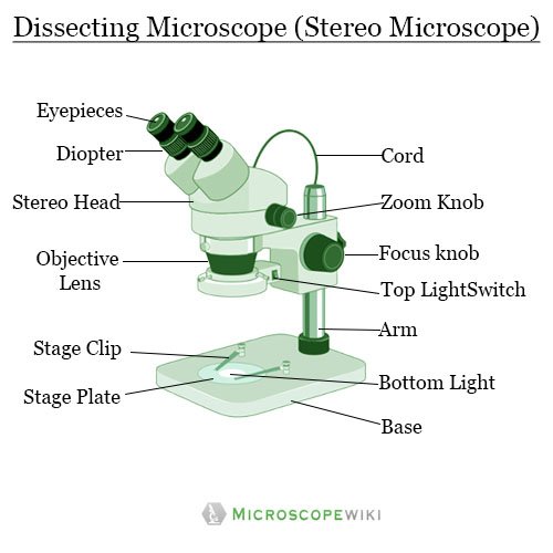
Parts of Stereo Microscope (Dissecting microscope ... Labeled part diagram of a stereo microscope Major structural parts of a stereo microscope Optical components of a stereo microscope - definition and function Eyepieces Eyepiece tube Diopter adjustment ring Interpupillary Adjustment Objective Lenses Barlow lens Adjustment Knobs Light sources Stage plate Stage chips Parathyroid Gland Histology with Microscope Slide Image ... The sample tissue section and diagram also show the numerous fat cells (adipose tissue). You may join anatomy learner on social media for a more updated labeled diagram on the parathyroid gland. Parathyroid gland microscope slide image drawing. This is a straightforward task to draw the microscope slide image of the parathyroid gland. Compound Microscope Parts - Labeled Diagram and their ... Labeled diagram of a compound microscope Major structural parts of a compound microscope There are three major structural parts of a compound microscope. The head includes the upper part of the microscope, which houses the most critical optical components, and the eyepiece tube of the microscope. Microscope Diagram Worksheet - The Microscope Create A ... Microscope Labeled Diagram from cdn.slidesharecdn.com Used to support the microscope when carried. There is a printable worksheet available for download here so you can take the . This online quiz is called microscope labeling game science, microsope. Be sure to check our teachers notebook store for other printables.
Simple Microscope - Parts, Functions, Diagram and Labelling ...
Label the microscope - Science Learning Hub Use this interactive to identify and label the main parts of a microscope . Drag and drop the text labels onto the microscope diagram. stage base diaphragm or iris light source eye piece lens high-power objective coarse focus adjustment fine focus adjustment Download Exercise Tweet

5 Important Types of Microscopes used in Biology (With Diagram)
Microscope labeled diagram - SlideShare Microscope labeled diagram 1. The Microscope Image courtesy of: Microscopehelp.com Basic rules to using the microscope 1. You should always carry a microscope with two hands, one on the arm and the other under the base. 2. You should always start on the lowest power objective lens and should always leave the microscope on the low power lens ...

Compound Microscope Parts, Functions, and Labeled Diagram ...
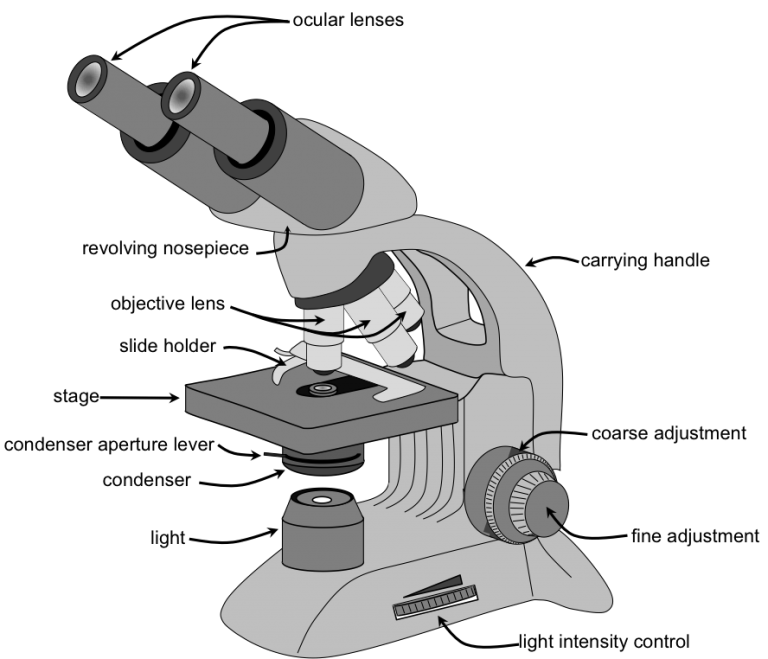
Parts of a microscope with functions and labeled diagram Figure: Diagram of parts of a microscope. There are three structural parts of the microscope i.e. head, base, and arm. Head - This is also known as the body, it carries the optical parts in the upper part of the microscope. Base - It acts as microscopes support. It also carries microscopic illuminators.

BIOLOGY FROM 1 | EQUIPMENTS USED FOR OBSERVATION | Cours ...
Compound Microscope - Diagram (Parts labelled), Principle ... Image : Labeled Diagram of compound microscope parts. See: Labeled Diagram showing differences between compound and simple microscope parts Structural Components. The three structural components include. 1. Head. This is the upper part of the microscope that houses the optical parts. 2. Arm . This part connects the head with the base and ...
Label a microscope - Teaching resources
Labeled Diagram Of A Stereo Microscope | Products ... Products/Services for Labeled Diagram Of A Stereo Microscope. Microscopes - (705 companies) Microscopes are instruments that produce magnified images of small objects Microscopes are instruments that produce a magnified image of a small object. They are used in many scientific and industrial applications. Some common applications...

Draw a well labelled diagram of a microscope. - Brainly.in
Microscope Types (with labeled diagrams) and Functions Phase-contrast microscope labeled diagram. Phase-contrast microscope functions: Its applications areas include. In cases where the specimen is colorless and is very tiny; In biology to conduct cellular level examination of microorganisms that can't be visualized using the bright field microscopy Interference Microscope

Free art print of Microscope
Diagram Of Animal Cell As Seen Under Light Microscope ... The diagram is very clear and labeled. Diagram of plant cell under microscope. Diagram Of Animal Cell Under Electron Microscope. A typical animal cell as seen in an electron microscope Medical Images For PowerPoint 1. But at the same time. Animal Cell Under Microscope. In such page we.
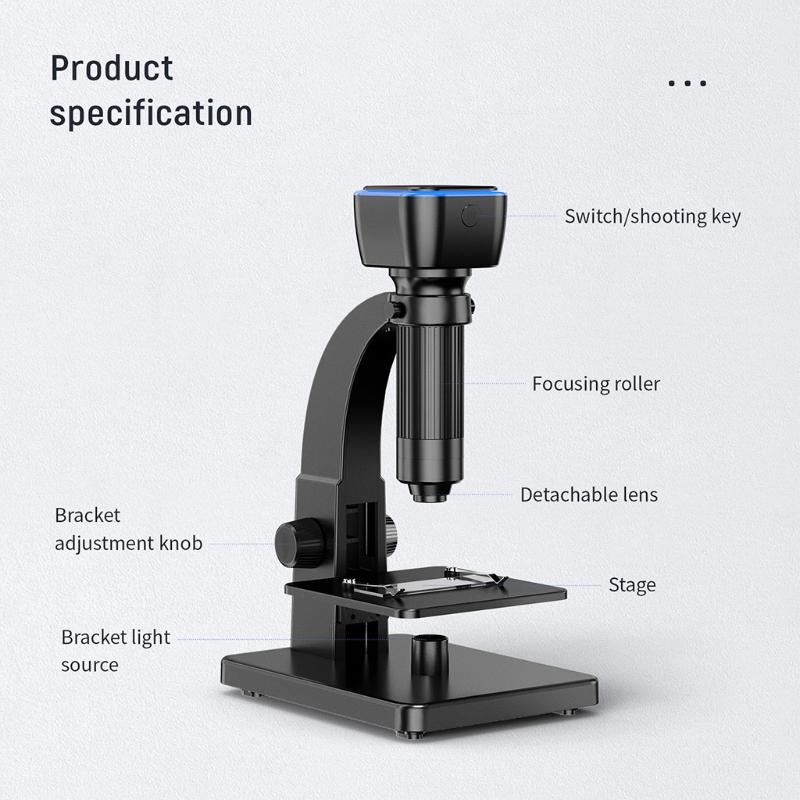
2000x Wifi Dual Lens Electronics Digital Microscope Usb Phone Repair Soldering Tool Industrial Sup
Compound Microscope Labeled Diagram | Quizlet Start studying Compound Microscope Labeled. Learn vocabulary, terms, and more with flashcards, games, and other study tools.

Microscope, Microscope Parts, Labeled Diagram, and Functions
Labelled Diagram Of A Plant Cell Under A Microscope ... Animal Cell Diagram Electron Microscope. 11 is a labelled diagram of a leaf palisade mesophyll cell as seen With a high quality light microscope. But at the same time. Under the microscope Priya observes a cell that has a cell wall and a distinct nucleus. Its a thin slice.

Compound Microscope: Definition, Parts, Application, Working ...
PDF Label parts of the Microscope: Answers Label parts of the Microscope: Answers Coarse Focus Fine Focus Eyepiece Arm Rack Stop Stage Clip . Created Date: 20150715115425Z ...
Mode d'emploi
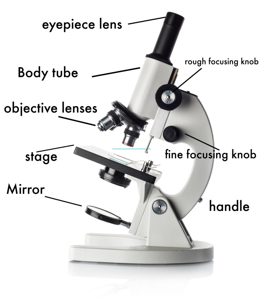
PDF Parts of the Light Microscope - Science Spot B. NOSEPIECE microscope when carried Holds the HIGH- and LOW- power objective LENSES; can be rotated to change MAGNIFICATION. Power = 10 x 4 = 40 Power = 10 x 10 = 100 Power = 10 x 40 = 400 What happens as the power of magnification increases?

Parts of Microscope | Function | Labeled Diagram | slidingmotion
Labelled Diagram of Compound Microscope - Biology Discussion The below mentioned article provides a labelled diagram of compound microscope. Part # 1. The Stand: The stand is made up of a heavy foot which carries a curved inclinable limb or arm bearing the body tube. The foot is generally horse shoe-shaped structure (Fig. 2) which rests on table top or any other surface on which the microscope in kept.
Draw a neat labeled ray diagram of a compound microscope ...
Compound Microscope Parts, Functions, and Labeled Diagram ... So, a compound microscope with a 10x eyepiece magnification looking through the 40x objective lens has a total magnification of 400x (10 x 40). Specimen or slide: The object used to hold the specimen in place along with slide covers for viewing. ... Compound Microscope Parts, Functions, and Labeled Diagram. Parts of a Compound Microscope.

Light Microscope- Definition, Principle, Types, Parts ...
Light Microscope- Definition, Principle, Types, Parts ... Figure: Labeled Diagram of a Light Microscope. Types of light microscopes (optical microscope) With the evolved field of Microbiology, the microscopes. used to view specimens are both simple and compound light microscopes, all using lenses. The difference is simple light microscopes use a single lens for magnification while compound lenses use ...

Microscope Parts and Functions
How to Use a Microscope
Solved Nikon Parts of the compound microscope Write the ...

Label the microscope — Science Learning Hub

Label Microscope Diagram - EnchantedLearning.com
Microscope PNG Collection d'images Téléchargement gratuit ...
(159).jpg)
Microscope Quiz: How Much You Know About Microscope Parts And ...

Compound Microscope Parts – Labeled Diagram and their ...

give a well labelled diagram of compound microscope using of ...

SWIFT Microscope élèves enfant SW150, 40X-1000X, tête ...

⭐️ MEILLEUR MICROSCOPE - Avis & Guide d'achat (Comparatif 2021)

label microscope diagram | Charts | Microscope, Polarizing ...

Parts of a microscope with functions and labeled diagram
22 Parts Of a Microscope With Their Function And Labeled ...

Parts of the Microscope with Labeling (also Free Printouts ...
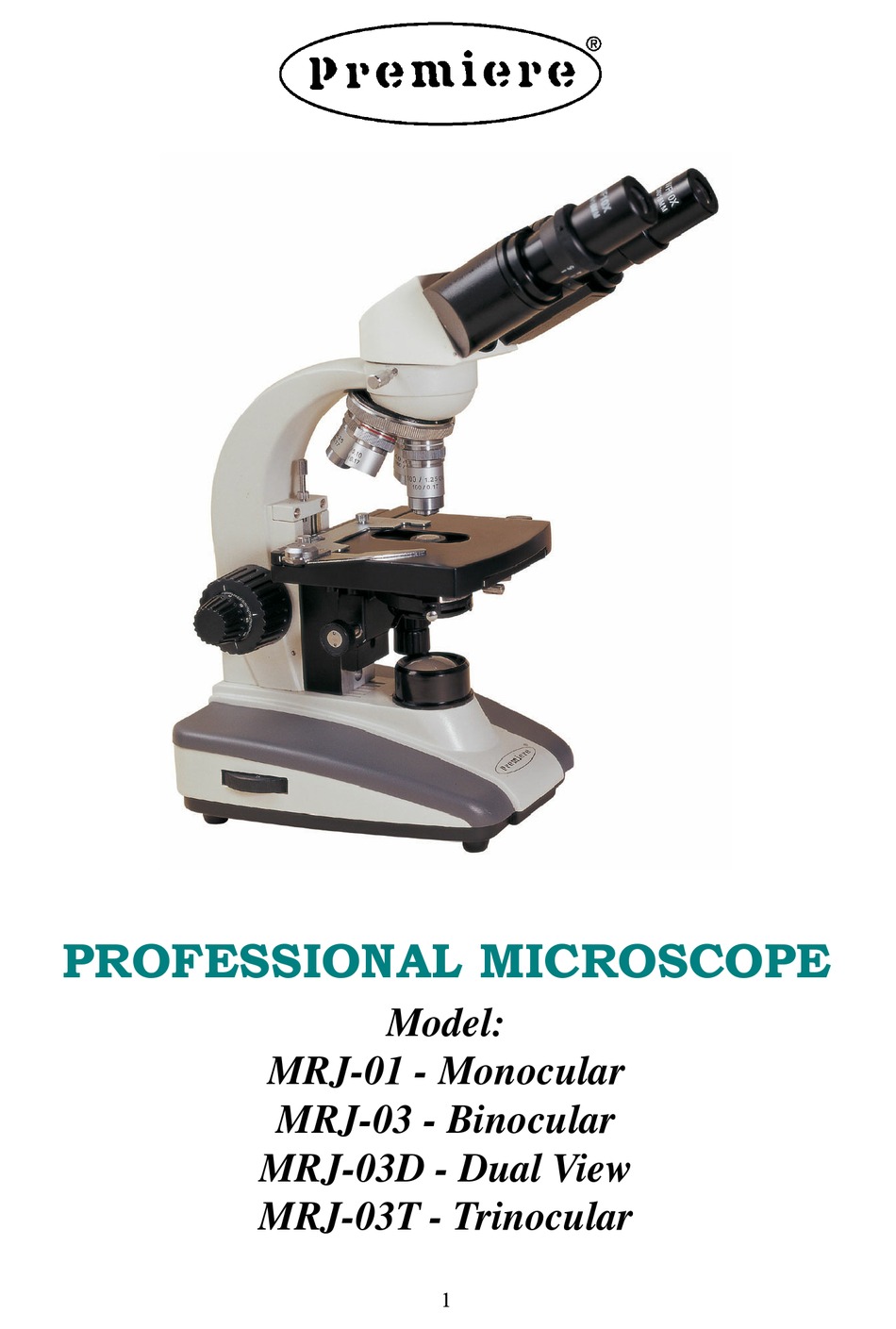
PREMIERE MRJ-01 MANUAL Pdf Download | ManualsLib

Draw a labelled diagram of a compound microscope.

MAXLAPTER Microscope Professionnel 100-1000x Platine ...

The Microscope
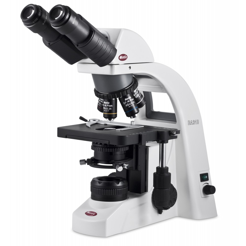
Microscope MOTIC BA310 Complet

Draw a neat labelled diagram of a compound microscope class ...
Microscopes Labeling Worksheets & Teaching Resources | TpT
Dissecting microscope (Stereoscopic or stereo microscope ...

Microscope optique - MF31-UV - Micro-shot Technology Limited ...
0 Response to "38 labeled diagram of a microscope"
Post a Comment